
Fluorescence in situ hybridisation (FISH) is an invaluable tool for the diagnostic and prognostic analysis of patient samples, providing a routine means of identifying genome duplications, deletions and rearrangements associated with disease.
The central component of any FISH assay is the probes. These are pools of fluorescently labelled DNA or RNA sequences designed to hybridise to target sequences, and that can subsequently be identified through imaging techniques such as fluorescent microscopy.
Discover how FISH can be used in cancer diagnostics and research
In situ probe design can be a challenging task for many labs, requiring significant time, money and expertise. However, without well-designed probes, your FISH experiments could be futile. But it’s not all doom and gloom, check out our five tips on what to look for when it comes the design of your in situ probes.
Before you spend time, effort, and expense designing and optimising probes for your FISH assay, it’s worth checking whether any suitable probes have already been designed and/or are commercially available. That way, you save precious time and money by minimising the validation process.
Suppliers, such as OGT, offer an extensive range of high quality, reliable and easy-to-use DNA FISH probes for many clinically-important applications. OGT’s CytoCell® probe range includes probes for the accurate detection of many haematological malignancies, common prenatal chromosomal disorders, and some of the rarest human genetic syndromes as well as the assessment of genetic aberrations in solid tumour samples.
Eight of OGT’s CytoCell FISH probes, designed to detect prenatal trisomy 13 & 21 and acquired cancer-related chromosome alterations, were recently granted IVDR certification, becoming the first FISH probes to achieve this. The IVDR certification means that customers can be confident in the continued quality, safety and performance of OGTs products and manufacturing processes.
Discover OGT’s range of CytoCell FISH probes and view our eight IVDR FISH probes
There are two main classes of probe that can be used for FISH: Probes based upon bacterial artificial chromosome (BAC) clones or oligo probes. BAC clones are distinct fragments of human chromosome that were originally cloned for sequencing as part of the Human Genome Project, but have since become highly valuable for FISH applications.
BAC clones can be substantial in size, averaging several hundred kilobases1, plus the entire BAC can be labelled with a fluorophore making for a relatively simple probe design workflow. In contrast, oligo probes are short, synthetic DNA fragments that are designed to specifically target a pre-determined region of interest.
Due to their significantly greater size, BAC probes offer greater coverage versus oligo probes. As such, BAC clones are able to target multiple genes or entire gene clusters, making them the ideal choice for detecting large genomic rearrangements, chromosomal aberrations and copy number changes. In light of the unparalleled coverage provided by BAC clone probes, all of OGT’s CytoCell FISH probes are based upon BAC clones.
Scientific papers are a valuable resource for in situ probe design, especially when searching for genetic aberration(s) associated with your disease of interest. By searching literature databases such as PubMed, you can find a wealth of study data highlighting clinically relevant genetic defects. From this, you can begin to compile a list of candidate aberrations to target with your FISH probes.
Once target aberrations have been identified, publicly available genomic databases such as Ensembl or UCSC can be leveraged to pinpoint BAC clones that could be included in your probe design. Examples of useful BAC clones could be those located at either side of a translocation breakpoint or a BAC clone inside a region that is known to be deleted or amplified.
Once you have carefully selected the appropriate BAC clones for your FISH assay, these can be purchased from BAC clone libraries or commercial vendors. It’s highly recommended to then validate the specificity of any BAC clones purchased by confirming their identity and integrity via sequencing and pilot FISH assays with control samples.
The myriad of fluorescent labels available for probe labelling can leave many of us feeling uncertain. There are many considerations to be made when selecting a fluorophore for your FISH probe, so here are some key factors to help you feel confident in your decision:
Depending on your application, how well studied your disease of interest is, and the level of expertise in your lab, FISH probe design and subsequent optimisation steps can take substantial time and effort. Expert third parties offer custom FISH probe design and manufacture services. For example, through its CytoCell myProbes® service, OGT experts can work with you to deliver optimised probes that meet your unique requirements – whether you need a simple modification of an existing catalogue product or require an entirely new and unique probe set.
Find out more about OGT’s CytoCell myProbes service
Since its inception, FISH technology has diversified. There are now countless different diagnostic tests in which scientists can exploit FISH and many different diseases and disorders can be studied. Therefore, while the tips and tricks above serve as a great platform for initial in situ probe design, each application will have its own nuances. Design guidelines will vary based on the particular assay you are using and the type of variation you’re targeting – but following these guidelines will provide a good start.
If you still need help designing your FISH probes, at OGT we’re here to help with our CytoCell myProbes® custom FISH probe design and manufacture service. Alternatively, why not see if your needs could be instantly met with our wide range of CytoCell FISH probes optimised for haematological malignancies and the assessment of genetic aberrations in solid tumour samples.
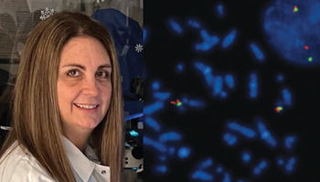
Tina Stafki talks about how she got hooked on FISH early on in her career, how she uses FISH in the CellNetix pathology lab to help patients and clinicians across the Pacific Northwest, and why CytoCell® FISH probes from OGT are her first choice when it comes to FISH probes.
Read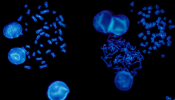
FISH is a cytogenetic technique utilised in labs to detect chromosomal abnormalities in both cancer and constitutional specimens. In this blog learn about the advantages of FISH...
Read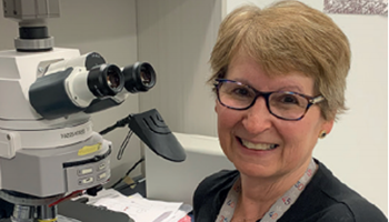
Carmen Vokaty, Assistant-Chief Medical Technologist in Cytogenetics at the McGill University Health Centre, details how her lab incorporated OGT’s Cytocell® Tissue Pretreatment kit into its workflow after struggling with a homebrew solution.
Read
Dr Jennie Thurston, Director of Cytogenetics, Carolinas Pathology Group, Atrium Health, explains how OGT's CytoCell® FISH probes were seamlessly incorporated into the automated workflow for fast, high quality results.
Read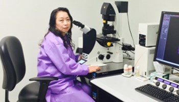
Megumi Hada, PhD, Senior Research Scientist, RaISE, Prairie View, Texas A&M. answers our questions on space radiation, how it affects astronaut health, and what her research can teach us about the biological effects of radiation here on Earth.
Read
Dr. Theresa C. Brown, Tulane University School of Medicine, answer questions around her AGT 2017 presentation 'FISHing with the real-life laboratory experts.'
Read Visit USA site
 Visit Canada site
