

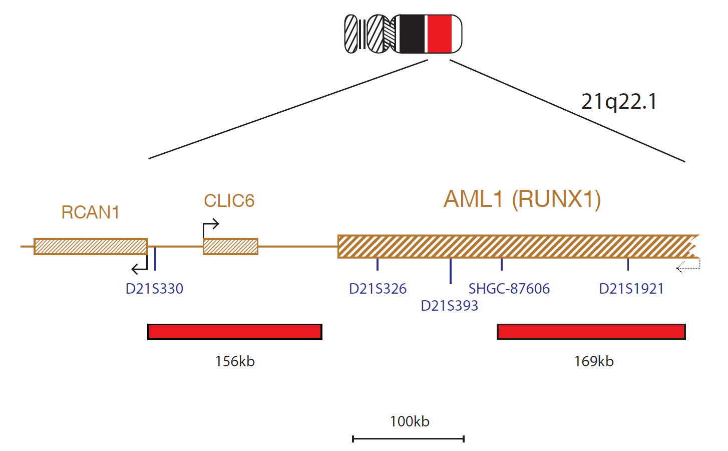
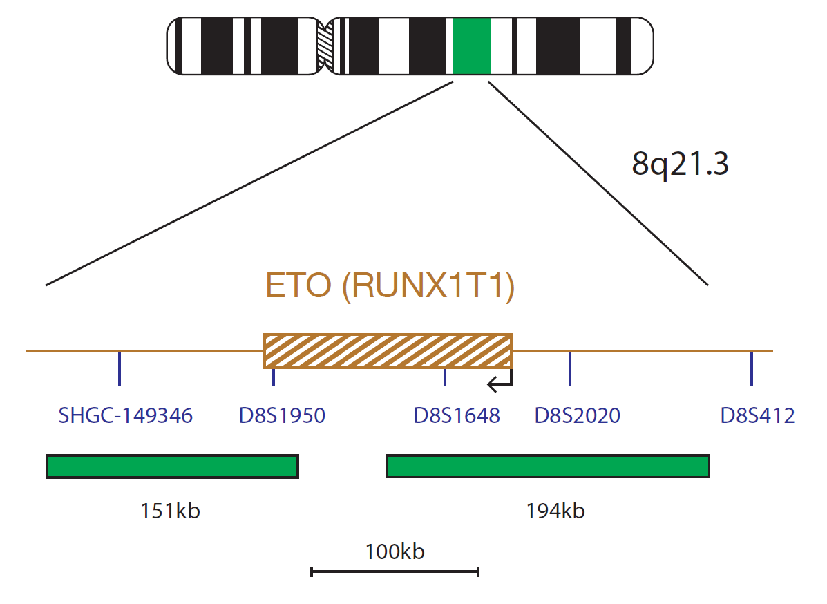
The AML1 component consists of a 156kb probe, labelled in red, located centromeric to the AML1 (RUNX1) gene that spans the CLIC6 gene and a 169kb probe covering part of the AML1 (RUNX1) gene, including markers SHGC-87606 and D21S1921. The ETO (RUNX1T1) component, labelled in green, consists of a 151kb probe covering the centromeric part of the gene and the flanking region and a 194kb probe covering the telomeric part of the gene and the flanking region.
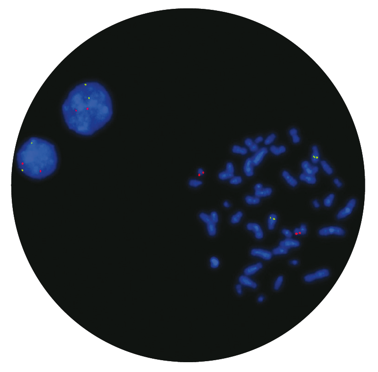
The RUNX1 (RUNX family transcription factor 1) gene at 21q22.12 is fused with the RUNX1T1 (RUNX1 partner transcriptional co-repressor 1) gene at Ensembl location 8q21.3, in the t(8;21)(q22;q22) translocation, found most commonly in patients with acute myeloid leukaemia (AML) FAB (French-American-British classification) type M2.
AML with a RUNX1-RUNX1T1 fusion resulting from a t(8;21)(q22;q22) translocation is a recognised disease entity according to the World Health Organization (WHO) classification of myeloid neoplasms and acute leukemia1. The translocation is observed in 10%-22% of patients with AML FAB type M2 and 5%-10% of AML cases overall, most commonly in children and young adults2 and is a good prognostic indicator3,4,5. The t(8;21) breakpoint mainly occurs in the intron between exons 5 and 6 just before the transactivation domain and fusion protein created contains the DNA-binding domain of RUNX1 fused to the transcription factor RUNX1T12.
In addition to the reciprocal t(8;21) translocation creating the RUNX1- RUNX1T1 fusion, variant translocations have also been reported. These variant rearrangements may be cryptic and easily overlooked by G-banding; however, FISH can indicate the presence of such rearrangements2.
Running our PETS protocol was taking upwards of 5 hours to complete based on the previous SOP. After the technical training visit from CytoCell, we were able to make some tweaks to reduce the protocol time down to just 1 hour and 15 minutes, with the same or better results.
Michelle Casey
Assistant Genetic Technologist, Leicestershire Genetics Centre, UK

 Visit USA site
 Visit Canada site


