


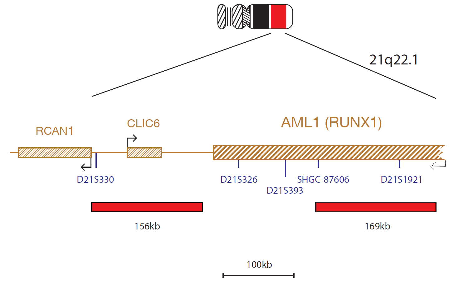
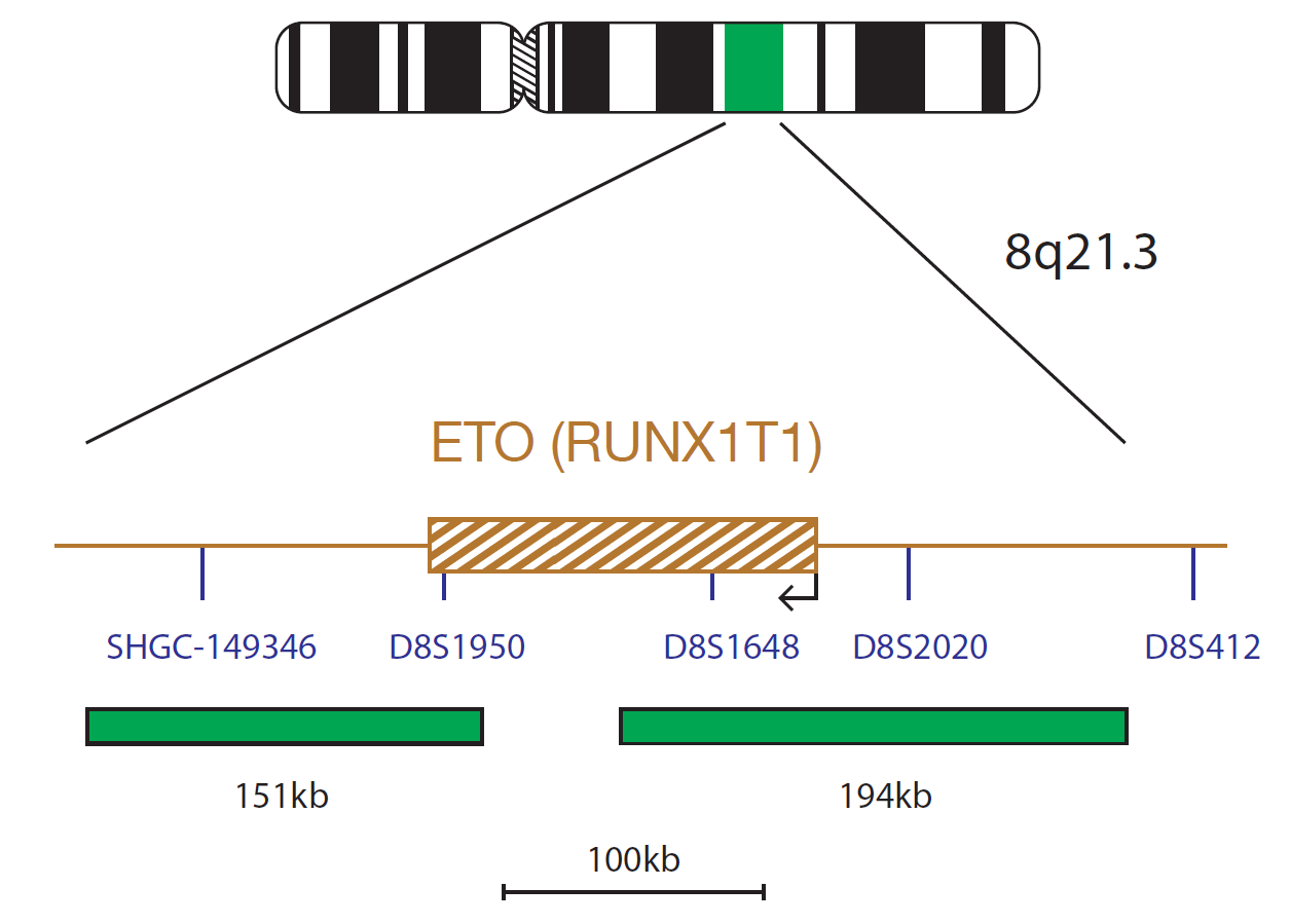
The AML1/ETO (RUNX1/RUNX1T1) Translocation, Dual Fusion FISH Probe Kit consists of a 156kb probe labeled in Texas Red, centromeric to the AML1 (RUNX1) gene, including the CLIC6 gene; a 169kb probe labelled in Texas red, telomeric to AML1 (RUNX1) gene, extending beyond the marker D21S1921; and two (151kb and 194kb) probes, labeled inn FITC green, on either side of the ETO (RUNX1T1) gene.
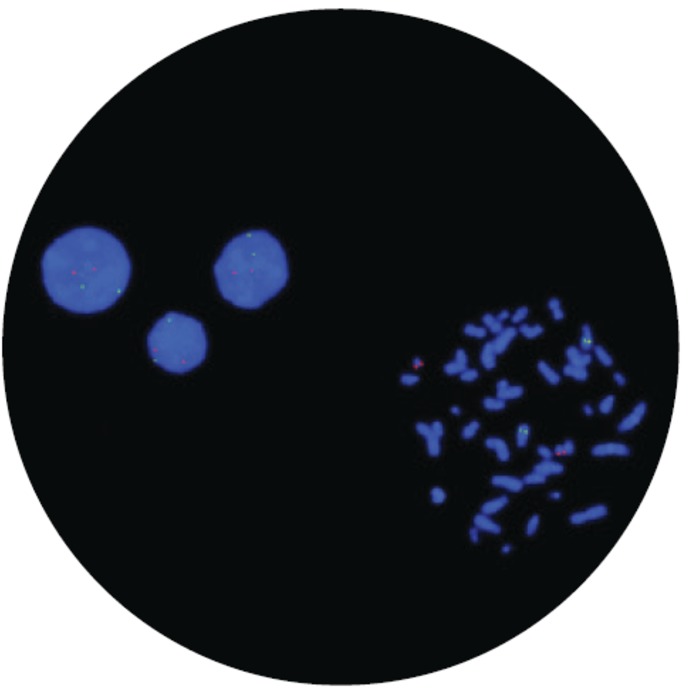
Microscope image
AML with a RUNX1-RUNX1T1 fusion resulting from a t(8;21)(q22;q22) translocation is a recognized disease entity according to the World Health Organization (WHO) classification of myeloid neoplasms and acute leukemia1. The translocation is commonly observed in patients with AML FAB type M2, most commonly in children and young adults2 and is a good prognostic indicator3,4,5. The t(8;21) breakpoint mainly occurs in the intron between exons 5 and 6, just before the transactivation domain. The fusion protein created contains the DNA-binding domain of RUNX1 fused to the transcription factor RUNX1T12. In addition to the reciprocal t(8;21) translocation creating the RUNX1-RUNX1T1 fusion, variant translocations have also been reported. These variant rearrangements may be cryptic and easily overlooked by G-banding; however, FISH can indicate the presence of such rearrangements2.
The AML1/ETO (RUNX1/RUNX1T1) Translocation, Dual Fusion FISH Probe Kit is a fluorescence in situ hybridization (FISH) Test used to detect rearrangement involving the AML1 (RUNX1) region on chromosome 21 at location 21q22.1 and the ETO (RUNX1T1) region on chromosome 8 at location 8q21.3 in fixed bone marrow specimens from patients with acute myeloid leukemia (AML). The test is indicated for characterization of patient specimens consistent with World Health Organization (WHO) guidelines for Classification of Tumours of Haematopoietic and Lymphoid Tissues (Revised 4th Edition) and in conjunction with other clinicopathological criteria. The assay results are intended to be interpreted by a qualified pathologist or cytogeneticist. The test is not intended for use as a stand-alone diagnostic, disease screening, or as a companion diagnostic.
For In Vitro Diagnostic Use. Rx only.
Reporting and interpretation of FISH results should be consistent with professional standards of practice and should take into consideration other clinical and diagnostic information. This kit is intended as an adjunct to other diagnostic laboratory tests and therapeutic action should not be initiated on the basis of the FISH result alone. Failure to adhere to the protocol may affect the performance and lead to false results.
Each lab is responsible for establishing their own cut-off values. Each laboratory should test sufficiently large number of samples to establish normal population distribution of the signal levels and to assign a cut-off value. The product is for professional use only and is intended to be interpreted by a qualified Pathologist or Cytogeneticist.
The device has not been specifically validated in patients with <20% blast count.
For sale in the US only. This product has not been licensed in accordance with Canadian law.
Find certificate of analysis documentation for our CytoCell FISH probes

In our hands, CytoCell FISH probes have proven to be of the highest quality with bright, easy to interpret signals, thus providing confidence in our results. OGT's customer support is outstanding, as their staff are extremely knowledgeable and truly care about their customers and their customers’ needs.

Jennie Thurston
Director of Cytogenetics, Carolinas Pathology Group, USA
I first came across CytoCell FISH probes in a previous lab I worked in and I was struck by the quality of the products. Since this time, I have been recommending and introducing CytoCell probes across all application areas — now they are the primary FISH probes used in our lab. They have an excellent range of products and their ready-to-use reagent format saves considerable time.

Elizabeth Benner
Medical Technologist, University of Arizona Health Network, USA
Our lab has been using a wide range of CytoCell FISH probes for a number of years, and have been increasing this range all the time. The probes have clear bright signals and show good reproducibility. CytoCell provides fast delivery of catalog probes, and are very responsive when we have any queries or problems with their products.

Bridget Manasse
Addenbrookes Hospital, Cambridge University Hosiptals NHS Foundation Trust, UK
The quality and consistency of CytoCell’s probes means I can trust the results, and my clients get their results in a timely manner.

Dr. Theresa C. Brown
Director, Cytogenetics Laboratory, Hayward Genetics Center, Tulane University School of Medicine, USA
It was very important for us to have more consistent results with our probes — easy-to-read bright signals and a range of vial sizes, which is much more cost-effective.

Janet Cowan, PhD
Director of the Cytogenetics Laboratory, Tufts Medical Center, USA
Not only do CytoCell offer an extensive range of high-quality FISH probes, the customer support is also excellent — providing fast access to all the probes I need. The probes are highly consistent with bright signals allowing easy scoring of results.
Dr. Eric Crawford
Senior Director, Genetics Associates Inc., USA
We have been working with CytoCell fish probes for two decades because of their excellent clarity and intensity regardless of the size of the probe. It is so clear and simple to detect.
Dr. Marina Djurisic
Head of Laboratory of Medical Genetics, Mother and Child Health Care Institute of Serbia “Dr Vukan Cupic”, Belgrade, Serbia
The quality and reproducibility of results using the CytoCell kit has been vital in accurately detecting co-deletions in our glioma investigations. We now have a cost-effective test that we can rely on that is also easy to use and interpret. We've been consistently impressed with this kit - not to mention the support offered by OGT's customer service, and have completely transitioned over to CytoCell probes.
Gavin Cuthbert, FRCPath
Head of Cancer Cytogenetics, Northern Genetics Servce, Newcastle, UK
 Visit International site
 Visit Canada site


