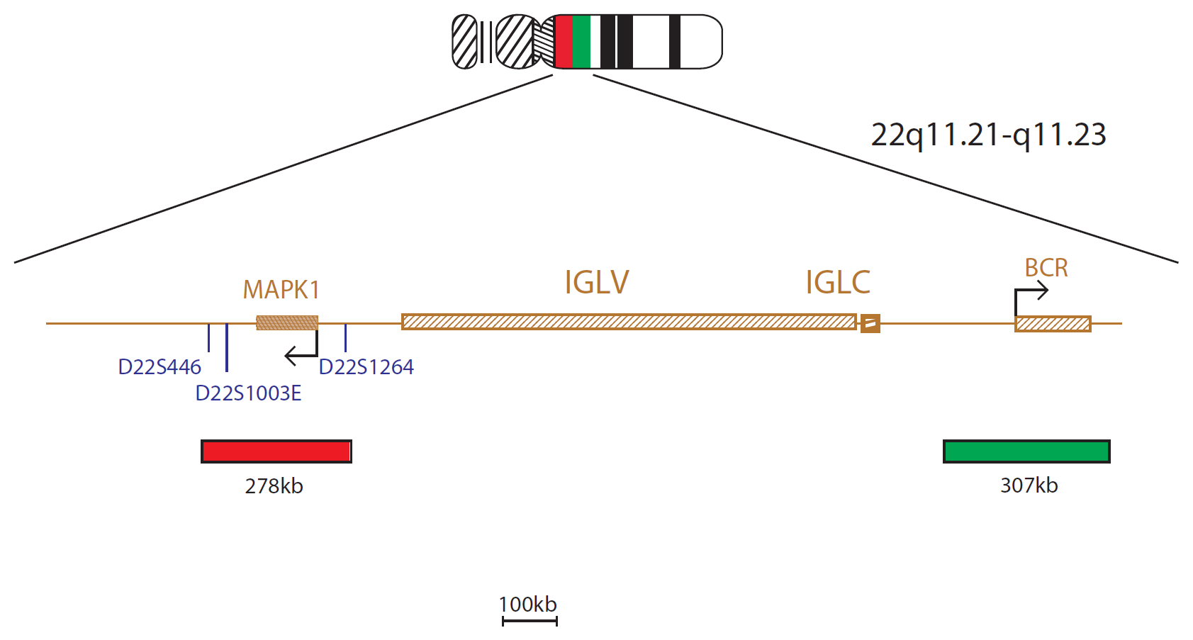
The IGL product consists of a 278kb probe, labelled in red, centromeric to the IGL Variable region and covering the MAPK1 gene, and a green probe, covering a 307kb region telomeric to the IGL Constant segment, including the BCR gene.
Translocations involving the immunoglobulin loci are recurring events in various subtypes of B-cell lymphomas.
In addition to translocations involving the IGH locus, variant translocations have been described in 5-10% of B-cell neoplasms involving either the immunoglobulin kappa (IGK) light chain locus at 2p11.2 or the lambda light chain (IGL) at 22q111,2. The best known translocations involving IG light chain loci are the variant Burkitt's translocations t(2;8)(p12;q24) and t(8;22)(q24;q11) present in up to 21% of all Burkitt's lymphomas3.
Other translocations involve the BCL6 oncogene, the t(2;3)(p12;q27) and t(3;22)(q27;q11) and BCL2 locus, t(2;18)(p12;q21) and t(18;22)(q21;q11)4,5. Translocations involving the IG light chain loci usually lead to breakage within the joining region of the respective locus2. IGL consists of 38 potentially active variable (IGLV) gene segments, 35 pseudogenes and seven IGL constant gene segments, each with a joining (J)-segment IGL (J-C)1.
The quality and reproducibility of results using the CytoCell kit has been vital in accurately detecting co-deletions in our glioma investigations. We now have a cost-effective test that we can rely on that is also easy to use and interpret. We've been consistently impressed with this kit - not to mention the support offered by OGT's customer service, and have completely transitioned over to CytoCell probes.
Gavin Cuthbert, FRCPath
Head of Cancer Cytogenetics, Northern Genetics Service, Newcastle, UK

 Visit USA site
 Visit Canada site