

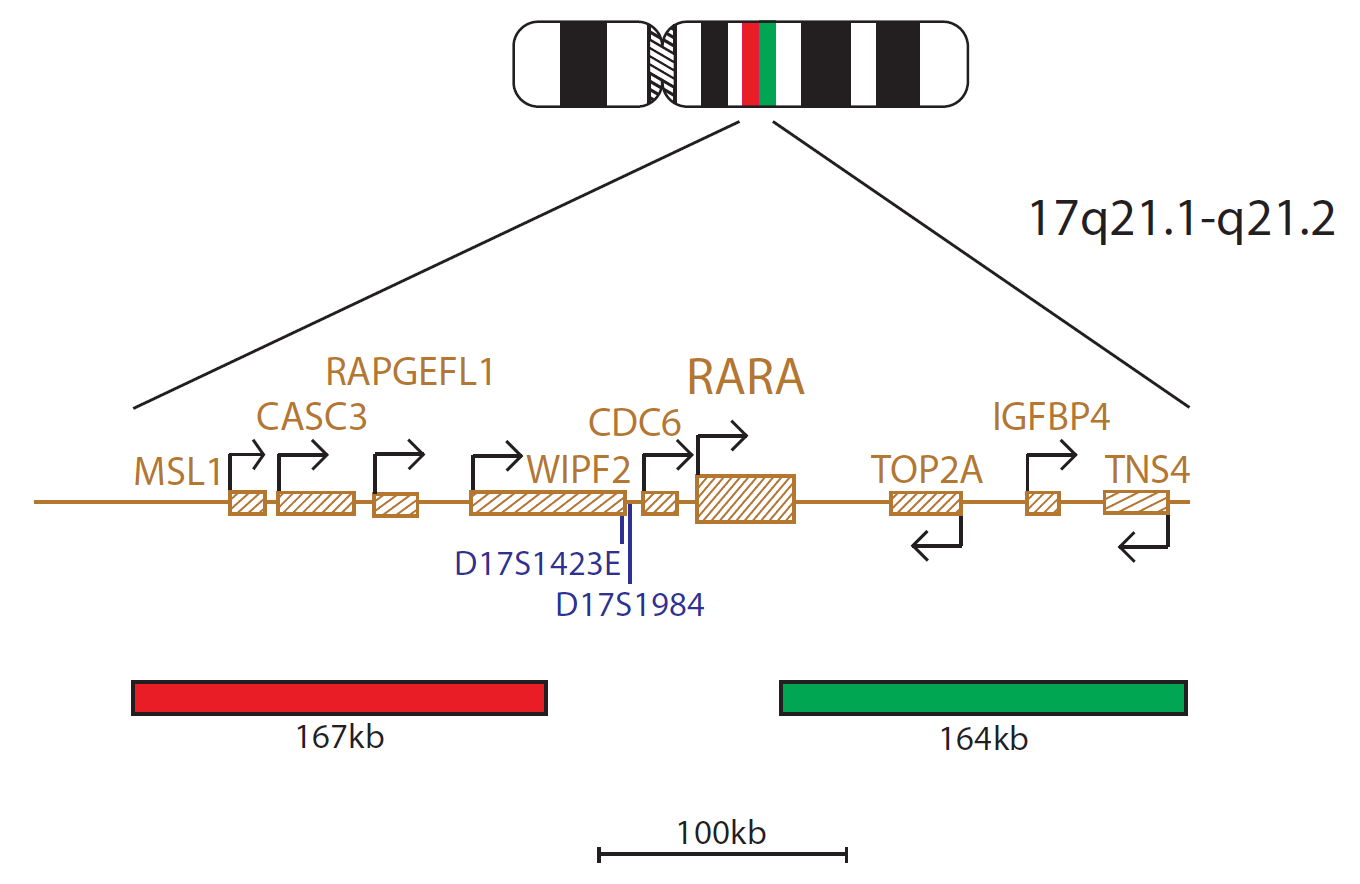
The RARα Breakapart probe mix consists of a 167kb probe centromeric to the RARα gene, including the CASC3 gene, and a 164kb probe, including the telomeric end of the RARα gene as well as the TOP2A and IGFBP4 genes.
The PML (promyelocytic leukemia) gene is located at 15q24.1 and the RARA (retinoic acid receptor alpha) gene is located at 17q21.2. In the vast majority of acute promyelocytic leukaemia (APL) cases, the RARA gene at 17q21.2 fuses with PML gene at 15q24.1; however, in <5% of cases of APL, RARA is fused to alternative partner1.
Known variant fusion partners include NPM1 at 5q35, NUMA1 at 11q13, ZBTB16 (PLZF) at 11q23, STAT5B at 17q21, PRKARIA at 17q24, FIP1L1 at 4q12 and BCOR at Xp111,2,3.
Patients with variant RARA translocations may show variable sensitivity to treatment with some patients showing resistance to treatment protocols1,3. It is therefore important to differentiate between APL patients with PML-RARA fusion and those patients with variant RARA translocations.
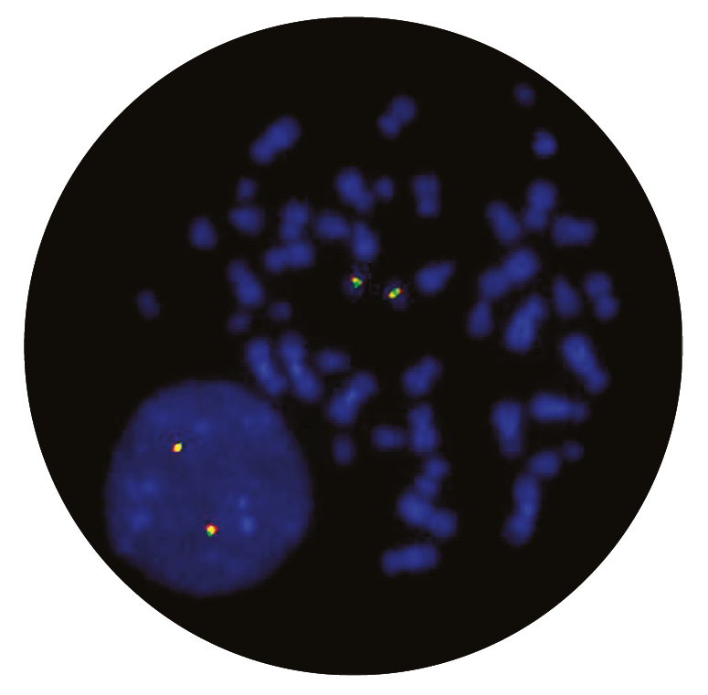
This breakapart probe will detect rearrangements of the RARA gene, irrespective of partner genes or chromosomes involved.
Running our PETS protocol was taking upwards of 5 hours to complete based on the previous SOP. After the technical training visit from CytoCell, we were able to make some tweaks to reduce the protocol time down to just 1 hour and 15 minutes, with the same or better results.

Michelle Casey
Assistant Genetic Technologist, Leicestershire Genetics Centre, UK

 Visit USA site
 Visit Canada site


