

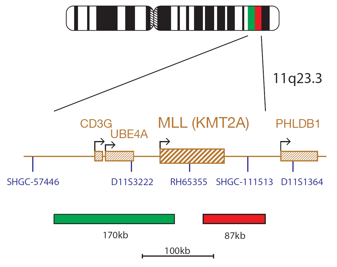
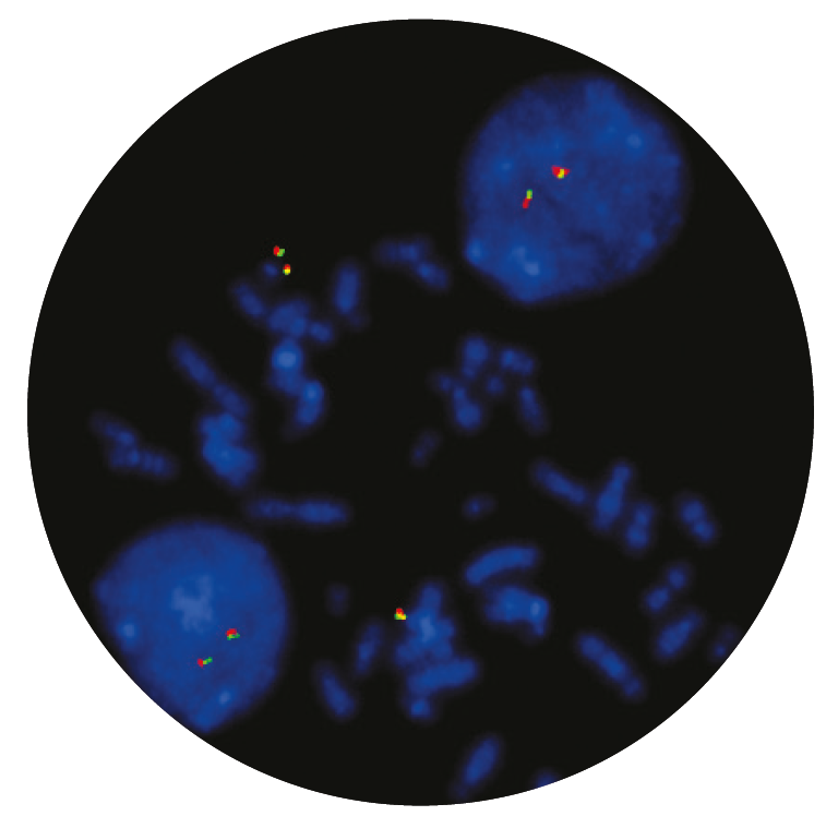
The MLL product consists of an 87kb probe, labelled in red, covering a region telomeric to the MLL (KMT2A) gene including the marker SHGC-111513 and a green probe covering a 170kb region centromeric to the MLL gene spanning the CD3G and UBE4A genes.
The KMT2A (lysine methyltransferase 2A) gene at 11q23.3 is commonly rearranged in acute leukaemias, especially in infant leukaemia and in secondary leukaemia, following treatment with DNA topoisomerase II inhibitors1.
The KMT2A gene has a great homology with the drosophila trithorax gene and encodes for a histone methyltransferase, which functions as an epigenetic regulator of transcription. KMT2A translocations result in the production of a chimeric protein in which the amino-terminal portion of KMT2A is fused to the carboxy-terminal portion of the fusion partner gene. The functional protein plays a critical role in embryonic development and haematopoiesis1,2,3,4.
KMT2A rearrangements can be detected in approximately 80% of infants with acute lymphoblastic leukaemia (ALL) and in 5-10% of paediatric and adult ALLs3,4. They can also be found in 60% of infant acute myeloid leukaemia (AML) and in 3% of de novo and 10% of therapy related adult AML cases3,5. To date, more than 70 partners have been identified with the most common translocations being MLL-AFF1; t(4;11) (q21;q23.3), MLL-MLLT4; t(6;11)(q27;q23.3), MLL-MLLT3;t (9;11) (p22;q23.3) and MLL-MLLT1; t(11;19)(q23.3;p13.3)1.
Running our PETS protocol was taking upwards of 5 hours to complete based on the previous SOP. After the technical training visit from CytoCell, we were able to make some tweaks to reduce the protocol time down to just 1 hour and 15 minutes, with the same or better results.

Michelle Casey
Assistant Genetic Technologist, Leicestershire Genetics Centre, UK

 Visit USA site
 Visit Canada site


