

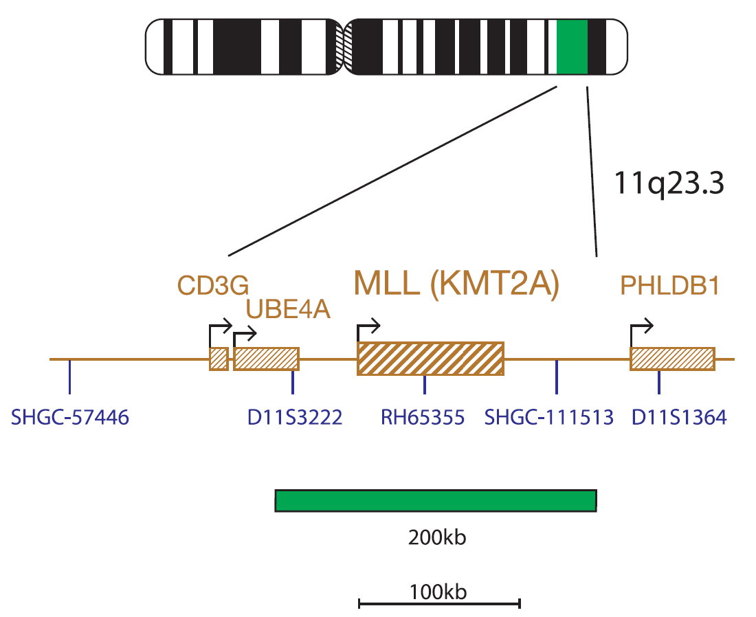
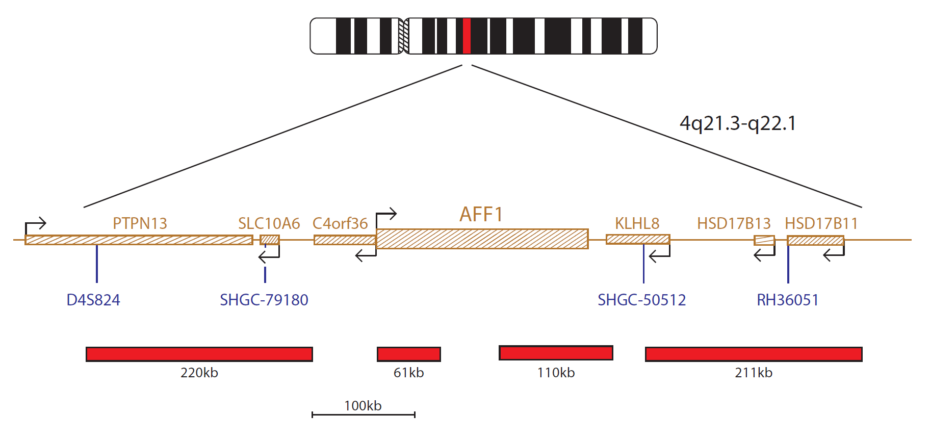
The MLL probe, labelled in green, covers a 200kb region including the MLL (KMT2A) gene. The AFF1 probe, labelled in red, consists of four clones (220kb, 61kb, 110kb and 211kb) covering the AFF1 gene and surrounding regions.
The KMT2A (lysine methyltransferase 2A) gene located at 11q23.3 and AFF1 (AF4/FMR2 family member 1) gene at 4q21.3 are involved in translocation t(4;11)(q21;q23.3), the most frequently observed translocation involving the KMT2A gene, in acute lymphoblastic leukaemia (ALL)1.
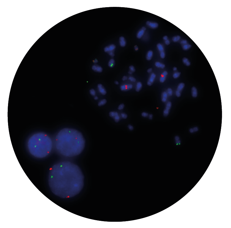
The t(4:11)(q21;q23.3) translocation results in the generation of two reciprocal fusion genes: KMT2A-AFF1 and AFF1-KMT2A – the leukaemic properties of the first have been documented but the role of the AFF1-KMT2A fusion protein is still under debate2,3,4.
UK best practice guidelines suggest that, if chromosome analysis is unsuccessful but FISH indicates a rearrangement of KMT2A, then further attempts to identify the t(4;11) must be made as the t(4;11)(q21;q23) is associated with a poor prognosis, and patients with this translocation may be treated on the high risk arm of MRC protocols5.
The MLL/AFF1 translocation, dual fusion probe allows both fusion genes, generated by the t(4;11)(q21;q23) translocation, to be detected.
Not only do CytoCell offer an extensive range of high-quality FISH probes, the customer support is also excellent — providing fast access to all the probes I need. The probes are highly consistent with bright signals allowing easy scoring of results.
Dr Eric Crawford
Senior Director, Genetics Associates Inc., USA

 Visit USA site
 Visit Canada site


