

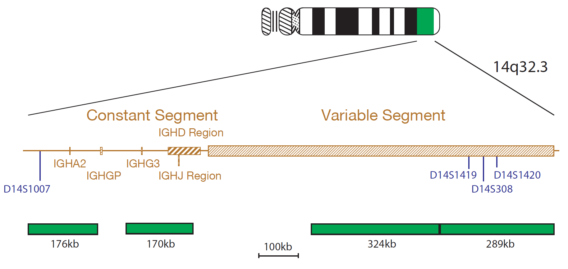
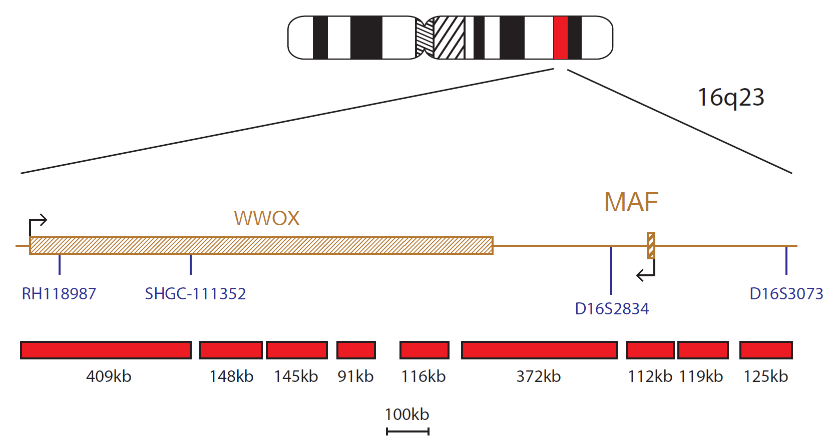
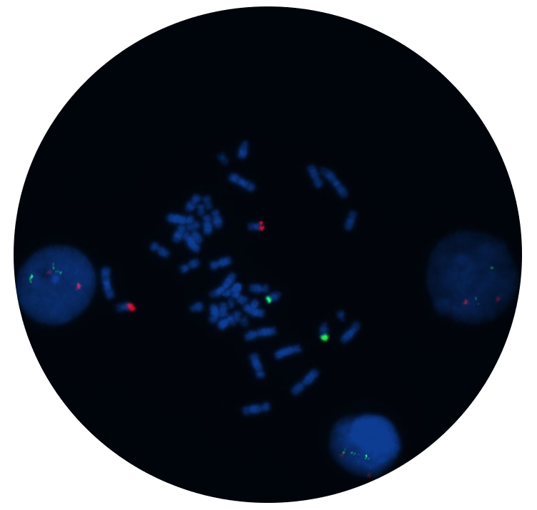
The IGH/MAF v2 Translocation, Dual Fusion Probe consists of the IGH probe mix, labelled in green, covering parts of the Constant, J, D and Variable segments of the IGH gene and the MAF probe mix, labelled in red, that encompasses the MAF gene and flanking regions as well as the WWOX gene.
MAF (MAF bZIP transcription factor) gene is located at 16q23 and IGH (immunoglobulin heavy locus) at 14q32.3. Approximately 50-60% of multiple myeloma (MM) cases are associated with translocations involving IGH and one of several partners including CCND1, NSD2 (WHSC1) and FGFR3, CCND3, MAF or MAFB1. The t(14;16)(q32.3;q23) translocation is a recurrent translocation seen in 2-10% of MMs1.
The majority of the breakpoints occur within the last intron of WWOX (WW domain containing oxidoreductase), centromeric to MAF. These breakpoints have a dual impact of positioning the IGH enhancer near MAF and disrupting the WWOX gene2. Gene expression profiling of myeloma cell lines revealed that MAF caused transactivation of cyclin D2 (a promoter of cell cycle progression), thus enhancing proliferation of myeloma cells3.
According to the literature, MM patients harbouring the t(14;16) appear to have a more aggressive clinical outcome4,5.
I first came across CytoCell FISH probes in a previous lab I worked in and I was struck by the quality of the products. Since this time, I have been recommending and introducing CytoCell probes across all application areas — now they are the primary FISH probes used in our lab. They have an excellent range of products and their ready-to-use reagent format saves considerable time.

Elizabeth Benner
Medical Technologist, University of Arizona Health Network, USA

 Visit USA site
 Visit Canada site


