

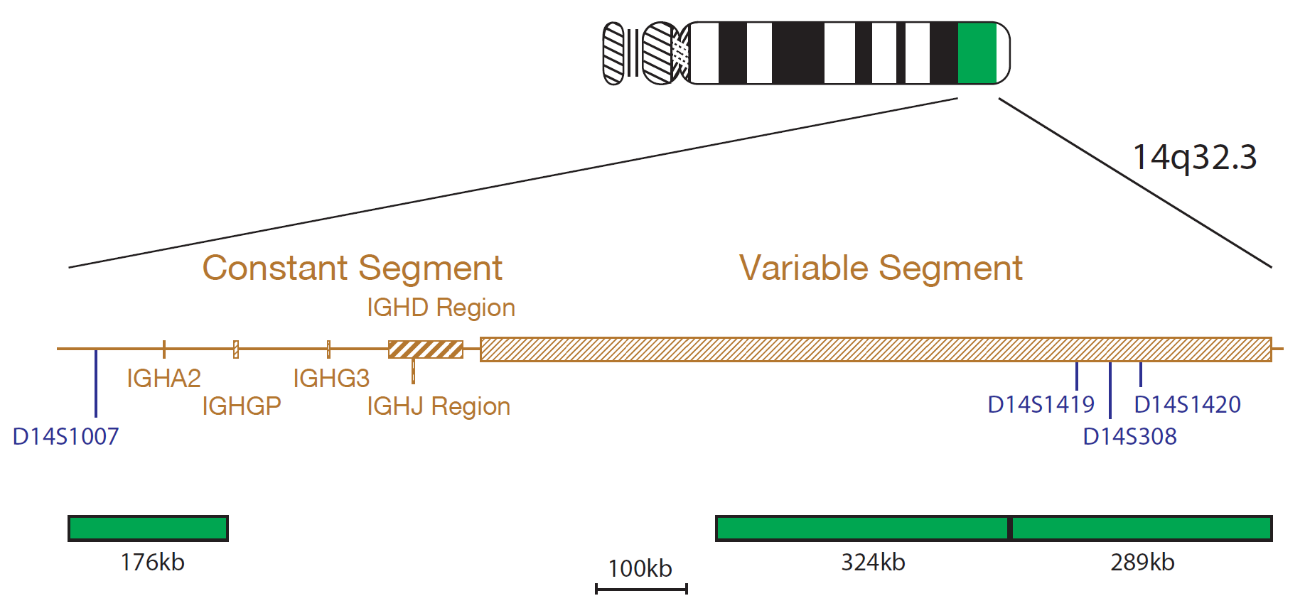
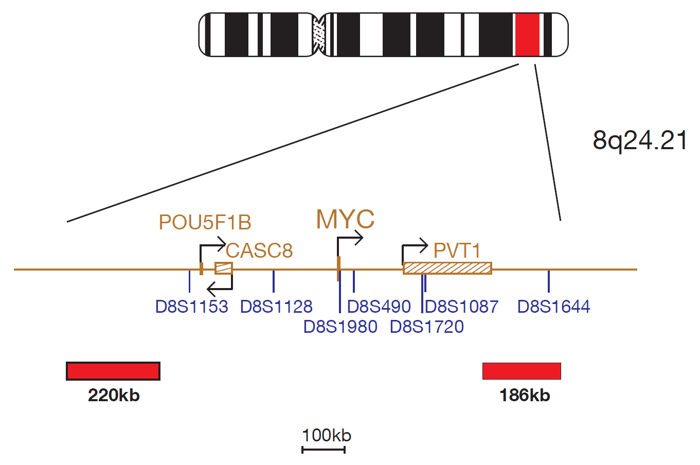
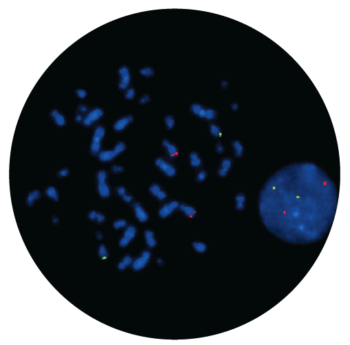
The IGH/cMYC product consists of probes, labelled in green, covering the Constant and Variable segments of the IGH gene, and cMYC probes, labelled in red. The cMYC probe mix contains a 220kb probe centromeric to the cMYC (MYC) gene and a second probe covering the 186kb region telomeric to the cMYC gene, including the D8S1644 marker.
The t(8;14)(q24;q32) translocation involving the IGH (immunoglobulin heavy locus) gene at 14q32.33 and the MYC (MYC proto-oncogene, bHLH transcription factor) oncogene at 8q24 is a recognised recurrent abnormality commonly seen in patients with B-cell malignancy.
IGH-MYC rearrangements are detected in up to 85% of cases of Burkitt lymphoma at diagnosis1. They are also seen in diffuse large B- cell lymphoma (DLBCL)2, multiple myeloma and plasmablastic lymphoma3,4.
In an IGH-MYC rearrangement the translocation breakpoints on chromosome 14 are clustered to a narrow region 5' to the intron enhancer of the immunoglobulin heavy chain, whereas the breakpoints on chromosome 8 can occur more than 340kb upstream of MYC, with no preferential site5. The translocation brings MYC into close proximity to the IGH enhancer and results in the up-regulation of MYC. Over-expression of the transcription factor stimulates gene amplification, resulting in uncontrolled cell proliferation6.
I am grateful for the excellent products I receive from CytoCell at a reasonable price, but more importantly the superb customer support. The speed in which I receive answers or suggestions makes my life as a director much easier and allows me to focus on patient care. The quality and consistency of CytoCell’s probes means I can trust the results, and my clients get their results in a timely manner

Dr. Theresa C. Brown
Director, Cytogenetics Laboratory, Hayward Genetics Center, Tulane University School of Medicine

 Visit USA site
 Visit Canada site


