

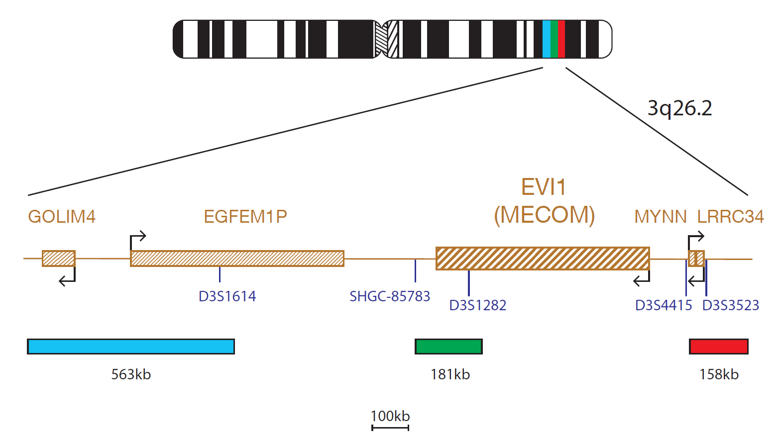
The red component of the EVI1 probe mix consists of a 158kb probe telomeric to the D3S4415 marker and includes the LRRC34 gene. The green component covers a 181kb region that includes the centromeric part of the EVI1 (MECOM) gene and beyond marker D3S1282. The blue component covers a 563kb region centromeric to the EVI1 gene, that includes the D3S1614 marker.
The MECOM (MDS1 and EVI1 complex locus) oncogene at 3q26.2 is often rearranged in haematological malignancies of myeloid origin.
MECOM encodes a zinc finger protein that is inappropriately expressed in the leukaemic cells of between 2-5% of AML and MDS patients1. This deregulated expression is often due to a chromosomal rearrangement involving 3q26.2, with the two most common aberrations being the t(3;3)(q21;q26.2) and inv(3)(q21q26.2)1. The breakpoints for the translocations and inversions vary considerably.
Inversion breakpoints are found centromeric to, and including, the MECOM gene and cover about 600kb. The majority of breakpoints in 3q26.2 translocations are telomeric to the MECOM gene and cover a region including the telomeric end of the MDS1 gene and the MYNN gene2.
Chromosome rearrangements involving the 3q26.2 region are associated with myeloid malignancies, aberrant expression of MECOM gene, an unfavourable prognosis and an aggressive clinical course2.
AML with inv(3)(q21q26.2) or t(3;3)(q21;q26.2) is a recognised disease entity according to the World Health Organization (WHO) classification of myeloid neoplasms and acute leukaemia. This is a transformed or de novo AML with a very aggressive clinical course and aberrations that involve MECOM at 3q26.2 and RPN1 (ribophorin I) at 3q213.
MECOM has also been shown to be rearranged in therapy-related disease via the t(3;21)(q26.2;q22) translocation, resulting in a MECOM- RUNX1 fusion3,4.
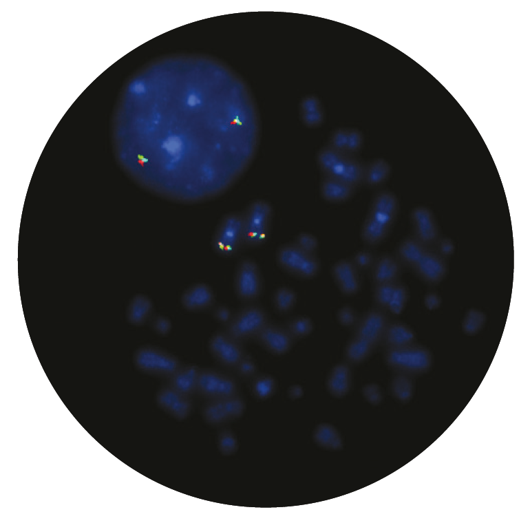
MECOM rearrangements are very heterogeneous and may be difficult to detect by conventional cytogenetics, making FISH a useful tool for their detection.
The quality of the products we have received from CytoCell have been excellent. The FISH probes they provide to us give intense, strong signals and are a pleasure to count. What has really stood out however has been the level of support and assistance provided by CytoCell’s application specialists. The team worked very closely alongside our own during the adoption of this product and spent many hours with us perfecting the technique, going above and beyond what I would expect during the transition period. Source BioScience absolutely demand high quality products and service to be able to deliver our results with confidence, and that is what we have received from CytoCell.
Neil Ryan
Laboratory Operations Manager, Source BioScience, UK

 Visit USA site
 Visit Canada site


