

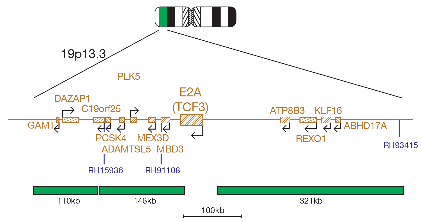
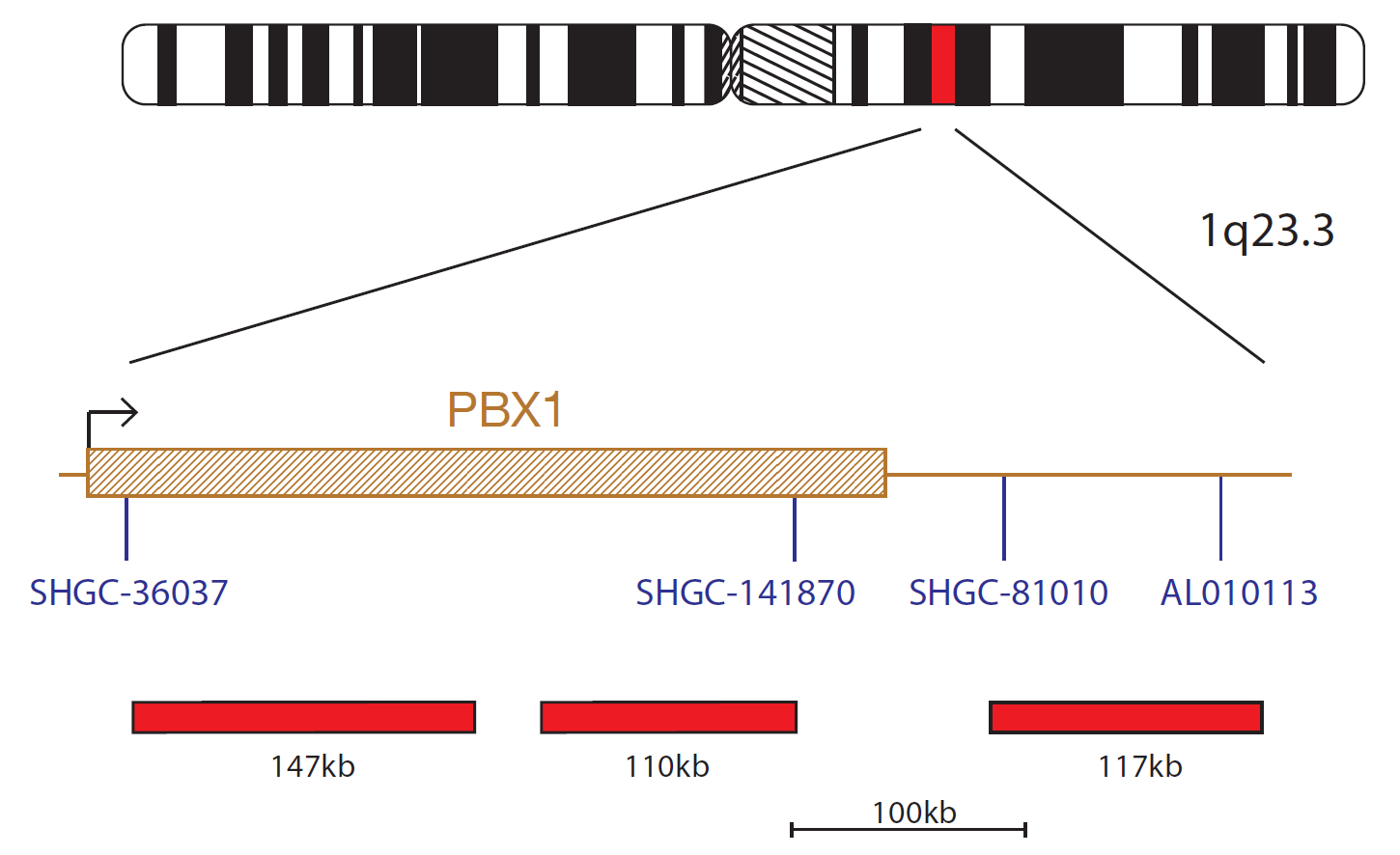
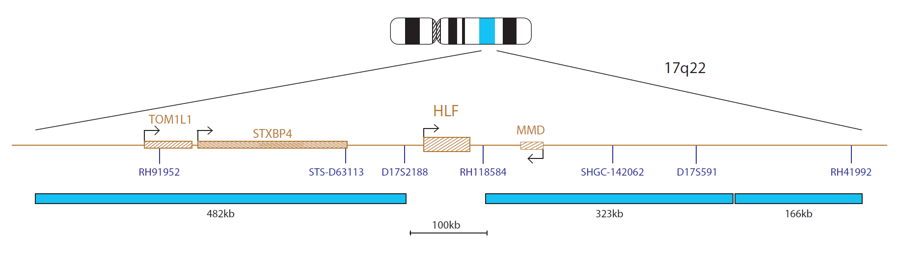
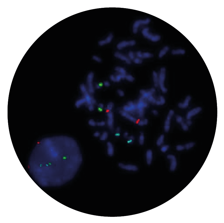
The E2A probe, labelled in green, contains two probes (110kb and 146kb) that cover the 3’ end of the E2A (TCF3) gene and flanking region and a 321kb probe that covers a region 5’ (centromeric) to the gene. The PBX1 probe, labelled in red, contains two probes (147kb and 110kb) that map within the PBX1 gene and a 117kb probe that maps 3’ (telomeric) to the gene. The HLF probe, labelled in blue, consists of a 482kb probe 5’ (centromeric) to HLF and two probes (323kb and 166kb) that are 3’ of the gene.
The TCF3 (transcription factor 3) gene is located at 19p13.3, PBX1 (pre-B-cell leukemia homeobox 1) gene is located at 1q23.3 and HLF (hepatic leukemia factor) at 17q22.
Translocations involving TCF3 are some of the most common rearrangements in childhood B-cell acute lymphoblastic leukaemia (ALL)1,2.
PBX1 and HLF become fused to TCF3 as a result of the t(1;19)(q23;p13) and t(17;19)(q22;p13) translocations, forming the TCF3-PBX1 and TCF3-HLF fusion genes, respectively. A rare cryptic inversion, inv(19)(p13;q13), has been reported to fuse TCF3 to TFPT (TCF3[E2A] fusion partner [in childhood leukaemia]), resulting in the TCF3-TFPT fusion gene1.
I first came across CytoCell FISH probes in a previous lab I worked in and I was struck by the quality of the products. Since this time, I have been recommending and introducing CytoCell probes across all application areas — now they are the primary FISH probes used in our lab. They have an excellent range of products and their ready-to-use reagent format saves considerable time.

Elizabeth Benner
Medical Technologist, University of Arizona Health Network, USA

 Visit USA site
 Visit Canada site


