

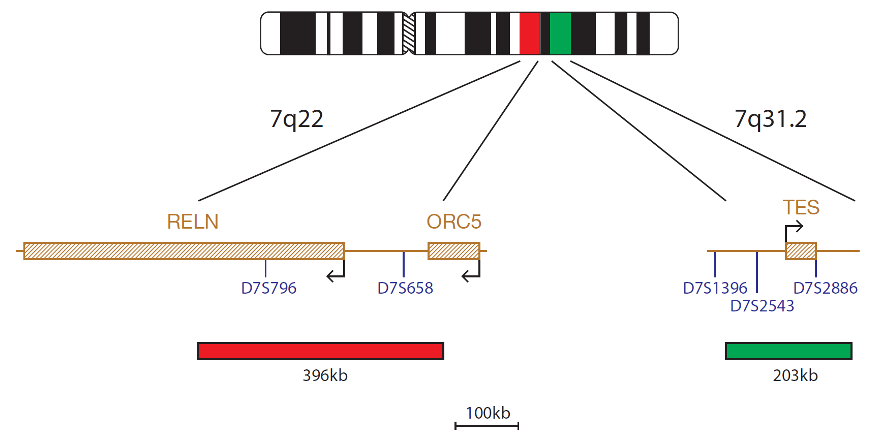
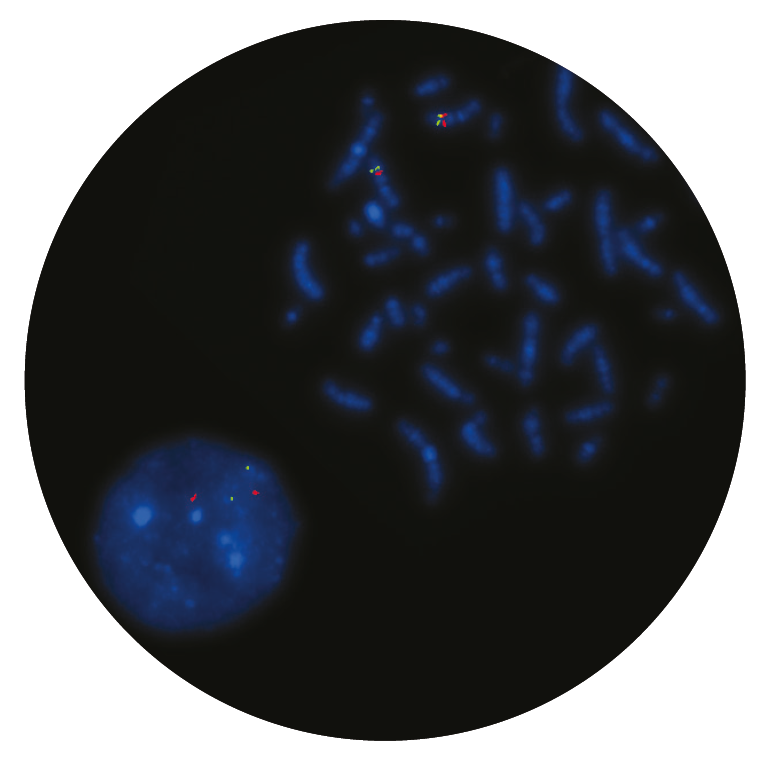
The 7q22 probe, labelled in red, covers a 396kb region including the telomeric end of the RELN gene and extending beyond the marker D7S658. The 7q31 probe, labelled in green, covers a 203kb region including the TES gene.
Monosomy of chromosome 7 and deletions of the long arm of chromosome 7 are recognised recurrent chromosomal aberrations frequently seen in myeloid disorders.
Monosomy 7 and del(7q) are seen in a number of myeloid disorders, including myelodysplastic syndrome (MDS), acute myeloid leukaemia (AML), and juvenile myelomonocytic leukemia (JMML)1. Furthermore, it occurs in MDS and AML that develop in patients with constitutional disorders (eg, Fanconi anaemia, Kostmann syndrome, neurofibromatosis type 1, and familial monosomy 7)2. The presence of Monosomy 7 or del(7q) as karyotypic changes are associated with a poorer outcome in myeloid malignancies1,3.
Deletions of chromosome 7 are typically large with heterogeneity in the breakpoints in myeloid diseases, making it difficult to map the common deleted regions (CDRs). It is highly likely that multiple tumour suppressor genes on chromosome 7 cooperate in leukaemogenesis4.
I am grateful for the excellent products I receive from CytoCell at a reasonable price, but more importantly the superb customer support. The speed in which I receive answers or suggestions makes my life as a director much easier and allows me to focus on patient care. The quality and consistency of CytoCell’s probes means I can trust the results, and my clients get their results in a timely manner

Dr. Theresa C. Brown
Director, Cytogenetics Laboratory, Hayward Genetics Center, Tulane University School of Medicine

 Visit USA site
 Visit Canada site


