

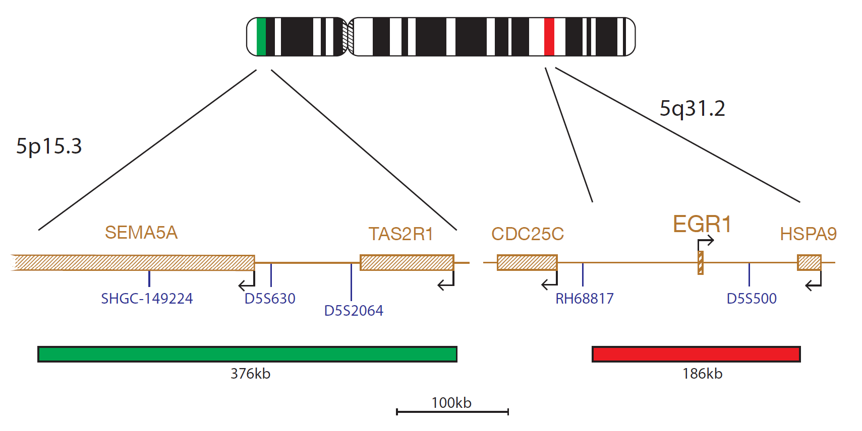
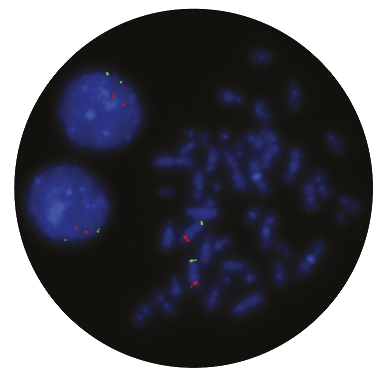
The EGR1 probe, labelled in red, covers a 186kb region within 5q31.2 that includes the D5S500 marker. The probe mix also contains a control probe, labelled in green for chromosome 5 at 5p15.3 that includes the marker D5S630.
Deletions of the long arm of chromosome 5 are one of the most common karyotypic abnormalities in myelodysplastic syndrome (MDS) and acute myeloid leukaemia (AML) with myelodysplasia related changes1,2.
A subset of patients with del(5q) as a sole cytogenetic abnormality has a consistent set of clinical features, termed the 5q- syndrome1. This clinical entity with <5% blasts has a more favourable prognosis and responds to treatment with lenalidomide. However, patients with del(5q) associated with other cytogenetic abnormalities or with excess blasts have an inferior survival2,3.
Two chromosomal regions have been mapped on chromosome 5q in MDS. One common deleted region, at 5q33, is associated with the 5q- syndrome. Another, more proximal region, located at 5q31, has been linked to a more aggressive form of MDS and AML and is often accompanied by additional cytogenetic abnormalities and a poorer prognosis1,3, 4.
The CytoCell® Del(5q) probe will detect deletions of EGR1 (early growth response 1), a tumour suppressor gene at 5q31. EGR1 has been shown to act through haploinsufficiency to initiate the development of MDS/AML5.
The quality of the products we have received from CytoCell have been excellent. The FISH probes they provide to us give intense, strong signals and are a pleasure to count. What has really stood out however has been the level of support and assistance provided by CytoCell’s application specialists. The team worked very closely alongside our own during the adoption of this product and spent many hours with us perfecting the technique, going above and beyond what I would expect during the transition period. Source BioScience absolutely demand high quality products and service to be able to deliver our results with confidence, and that is what we have received from CytoCell.
Neil Ryan
Laboratory Operations Manager, Source BioScience, UK

 Visit USA site
 Visit Canada site


