

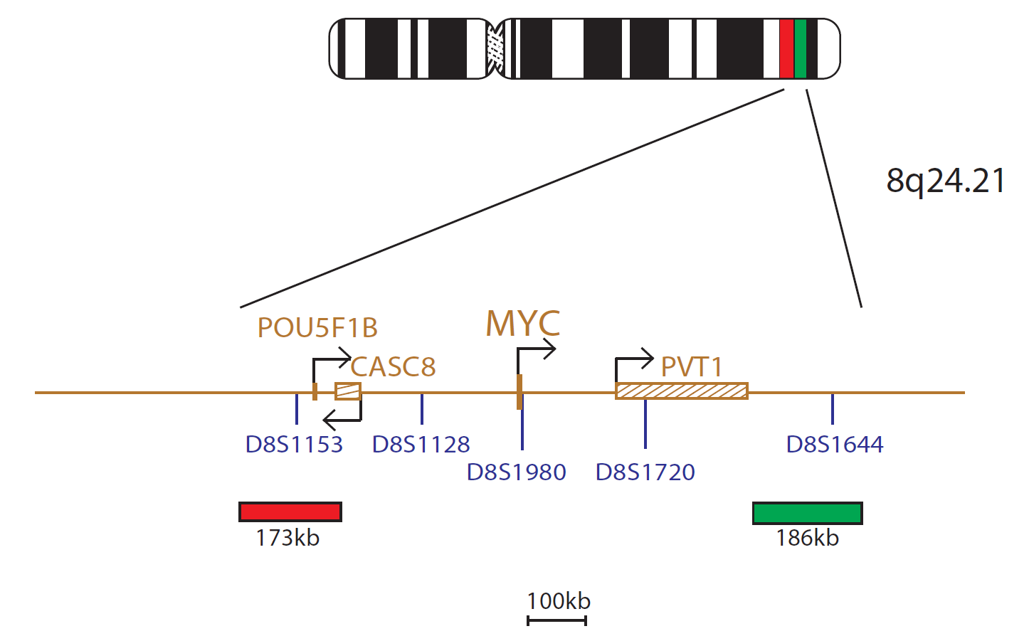
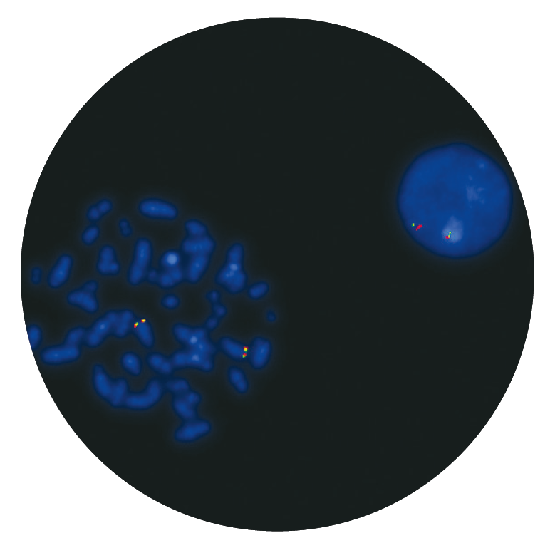
The cMYC probe mix consists of a 173kb probe labelled in red, centromeric to the MYC gene that includes the marker D8S1153 and a 186kb probe, labelled in green telomeric to the MYC gene that includes the D8S1644 marker.
Chromosomal rearrangements involving the MYC (MYC proto-oncogene, bHLH transcription factor) gene at 8q24 are recognised recurrent abnormalities commonly seen in patients with B-cell malignancy.
MYC rearrangements, activating MYC by translocation with one of the three immunoglobulin loci (IGH, IGL or IGK), are detected in almost all cases of Burkitt lymphoma at diagnosis1. They are also seen in diffuse large B-cell lymphoma (DLBCL)2, multiple myeloma and plasmablastic lymphomas3,4, amongst other diseases.
MYC has also been shown on rare occasions to be involved in rearrangements with a number of non-immunoglobulin partners5.
The presence of concurrent MYC rearrangements with BCL2 and/or BCL6 rearrangements in patients with ‘dual-hit’ lymphoma has been shown to be associated with aggressive disease6.
Running our PETS protocol was taking upwards of 5 hours to complete based on the previous SOP. After the technical training visit from CytoCell, we were able to make some tweaks to reduce the protocol time down to just 1 hour and 15 minutes, with the same or better results.

Michelle Casey
Assistant Genetic Technologist, Leicestershire Genetics Centre, UK

 Visit USA site
 Visit Canada site


