

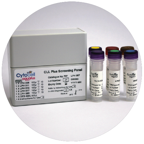
A selection of haematology probes and an alpha-satellite probe for Chronic Lymphocytic Leukaemia (CLL).
Probe specification
The Alpha Satellite 12 Plus Probe is a repeat sequence probe, labelled in red, which recognises the centromeric repeat sequence D12Z3.
Probe information
Trisomy 12 is a recurrent abnormality in CLL, seen in 20% of cases1 and often appears as the unique cytogenetic aberration (40-60% of cases with trisomy 12)2. Patients with trisomy 12 are classified as low-risk in the absence of any other genetic lesions3. This product is also available in 5 (LPH 069-S) and 10 (LPH 069) test kit sizes and has been optimised for overnight hybridisation.
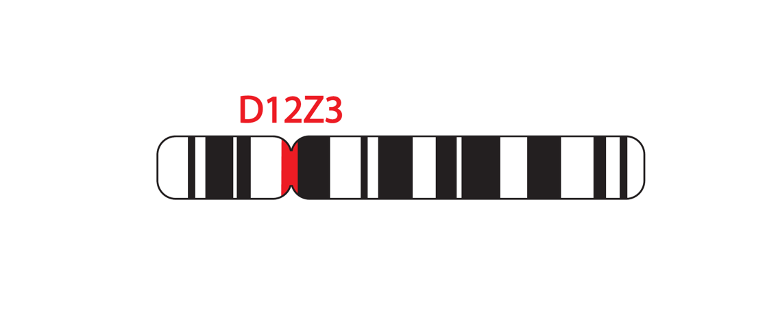
12p11.1-q11.1 (D12Z3)
Probe specification
The 13q14.2-q14.3 probe, labelled in red, covers the D13S319 and D13S25 markers. The 13qter subtelomere specific probe (clone 163C9), labelled in green, allows identification of chromosome 13 and acts as a control probe.
Probe information
Deletions affecting 13q14 are the most frequent structural genetic aberrations in CLL3,4,5. This region is found to be heterozygously deleted in 30-60% and homozygously deleted in 10-20% of CLL patients6. Patients with 13q14 deletions are classified as very low risk, in the absence of any other genetic lesions3.
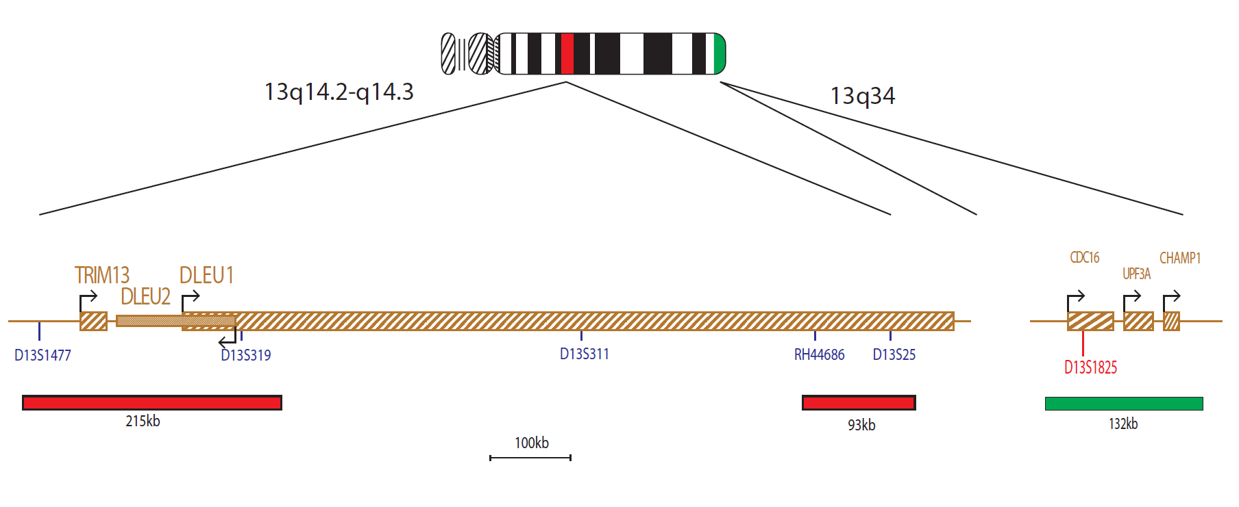
13q14.2-q14.3
Probe specification
The P53 probe mix consists of a 161kb probe, labelled in red that covers the whole P53 (TP53) gene and the flanking regions. The probe mix also contains a control probe for the 17 centromere (D17Z1) that is labelled in green.
Probe information
The TP53 (tumor protein p53) gene at 17p13 is one of most important tumour suppressor genes; it acts as a potent transcription factor with fundamental role in the maintenance of genetic stability. Loss of TP53 is reported in 10% of patients with CLL, and is considered to be the poorest prognostic marker3,7.
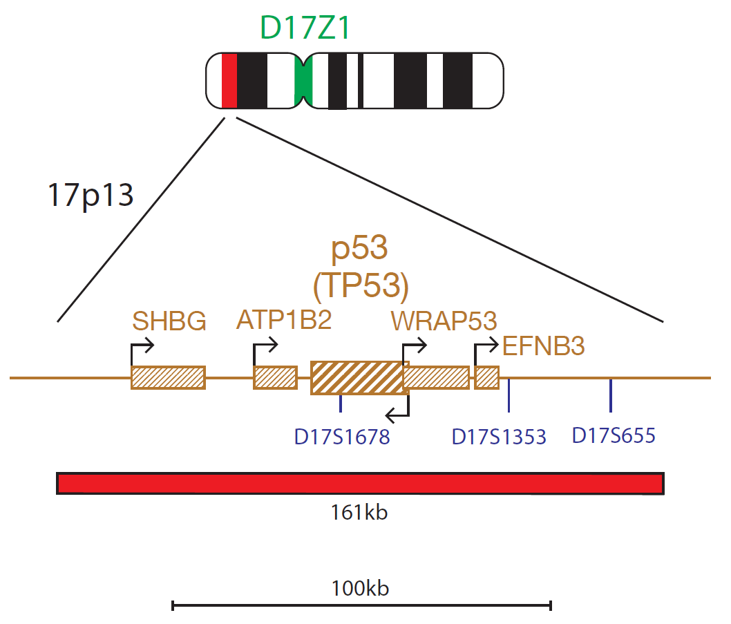
17p13.1 (P53)
Probe specification
The ATM probe is 182kb, labelled in red, and covers the telomeric end of the NPAT gene and the centromeric end of the ATM gene to just beyond the D11S3347 marker. The probe mix also contains a control probe for the 11 centromere (D11Z1) labelled in green.
Probe information
The ATM (ATM serine/threonine kinase) gene at 11q22.3 is an important checkpoint gene involved in the management of cell damage and its function is to assess the level of DNA damage in the cell and attempt repair by phosphorylating key substrates involved in the DNA damage response pathway8. Loss of ATM is reported in 18% of patients with CLL, and is considered a poor prognostic marker in CLL9.
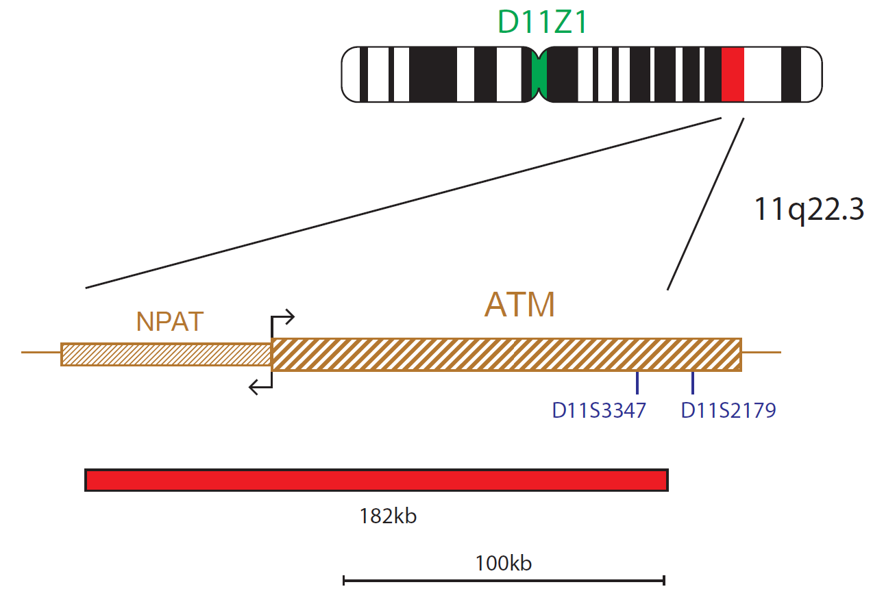
11q22.3 (ATM)
Probe specification
The MYB probe mix consists of a 183kb probe, labelled in red that covers the entire MYB gene and a region telomeric to the gene that includes a centromeric part of the AHI1 gene. This probe mix also contains a control probe for the 6 centromere (D6Z1) labelled in green.
Probe information
Deletions of chromosome 6q are recurrent in CLL. The MYB (MYB protooncogene, transcription factor) gene is essential in haematopoietic cell proliferation and differentiation10,11. It is located in band 6q23.3 and is provided as a marker for 6q deletion.
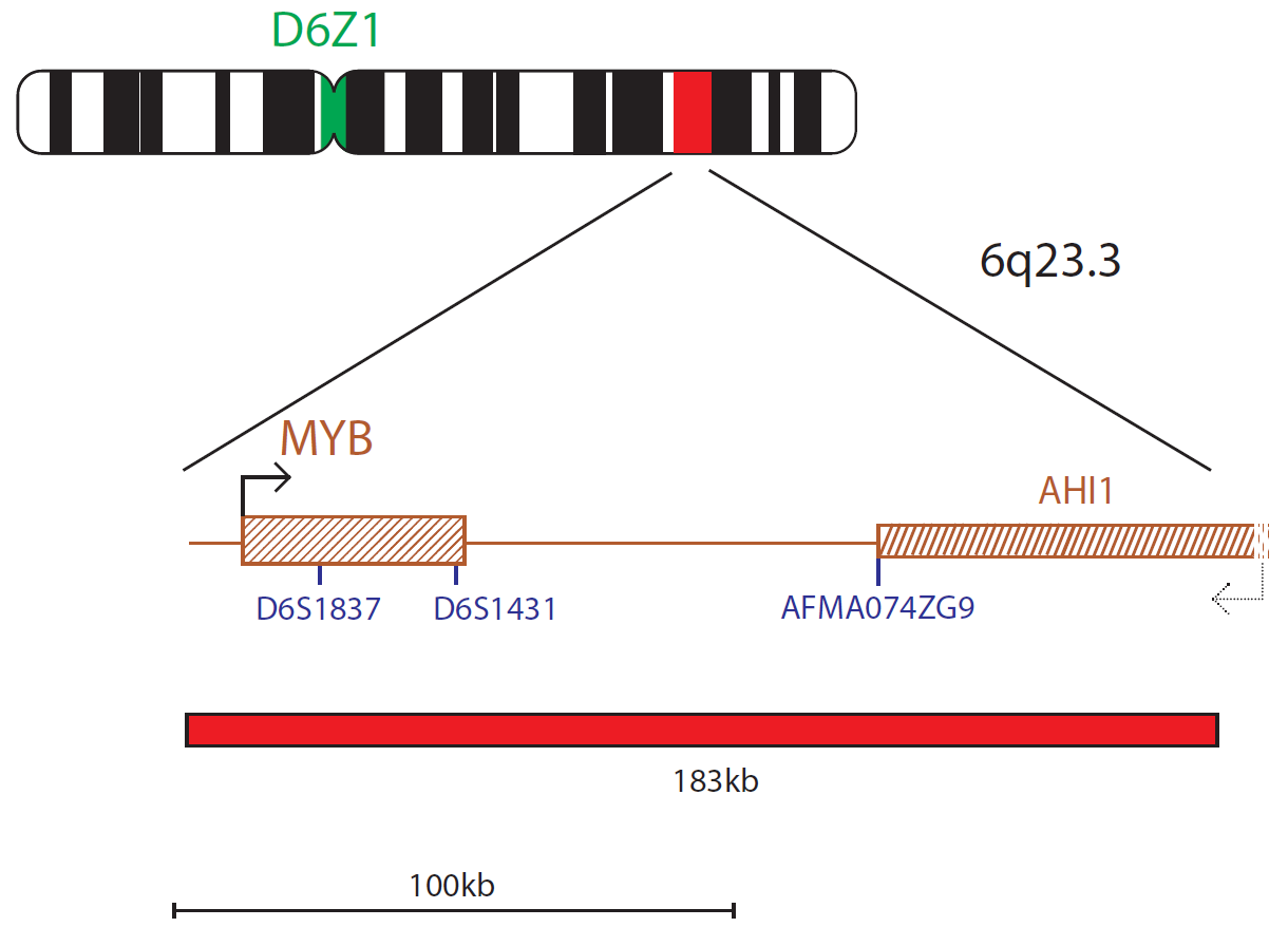
6q23.3 (MYB)
I first came across CytoCell FISH probes in a previous lab I worked in and I was struck by the quality of the products. Since this time, I have been recommending and introducing CytoCell probes across all application areas — now they are the primary FISH probes used in our lab. They have an excellent range of products and their ready-to-use reagent format saves considerable time.

Elizabeth Benner
Medical Technologist, University of Arizona Health Network, USA

 Visit USA site
 Visit Canada site


