

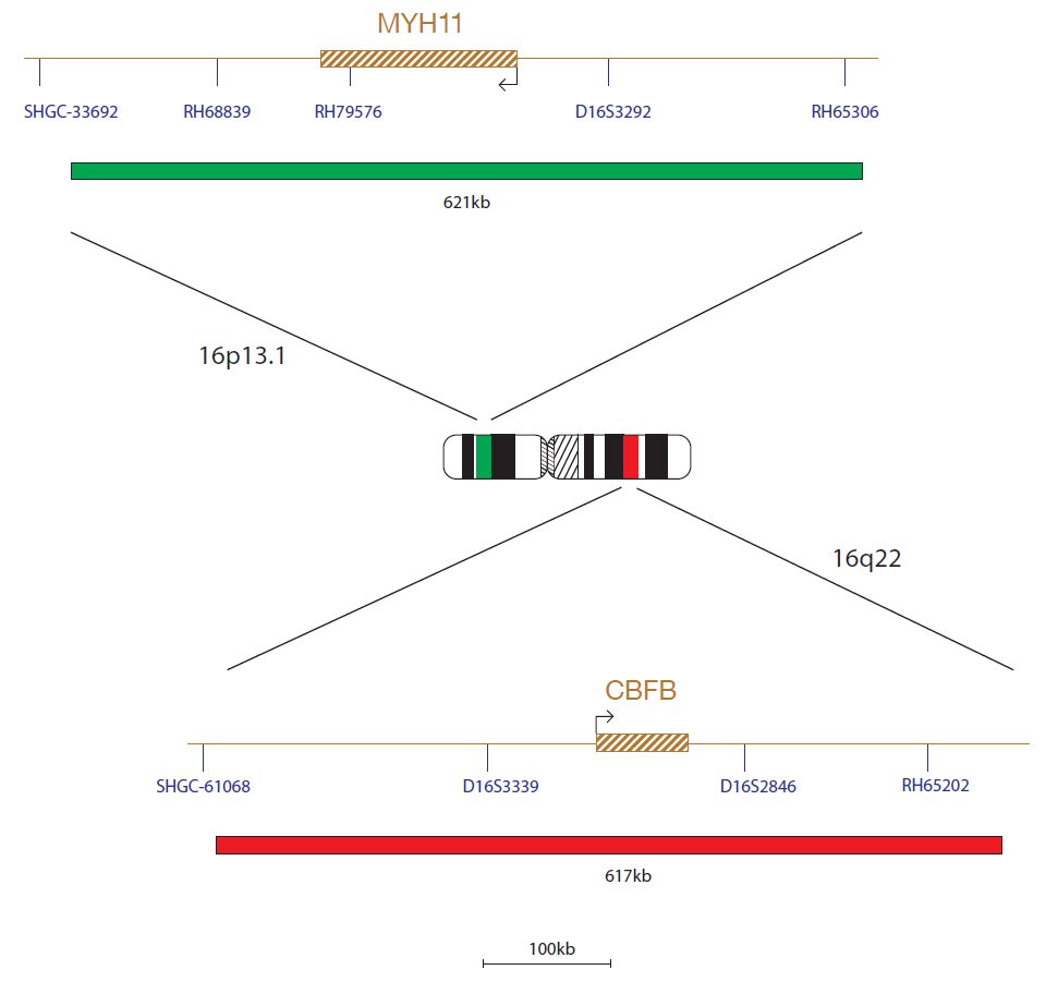
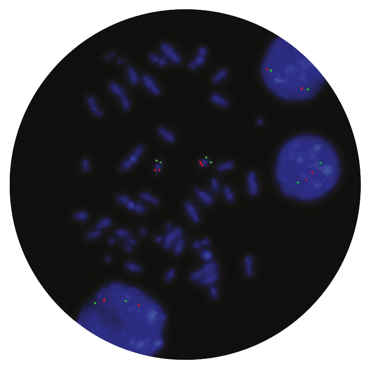
The CBFβ probe, labelled in red, covers a 617kb region, within 16q22.1 and includes the CBFβ gene. The MYH11 probe, labelled in green, covers a 621kb region within 16p13.11 and includes the MYH11 gene.
The CBFβ (core-binding factor subunit beta) gene is located at 16q22.1 and the MYH11 (myosin heavy chain 11) gene is located at 16p13.11. The inversion inv(16)(p13.11q22.1) and the translocation t(16;16) (p13.11;q22.1) give rise to the CBFβ-MYH11 fusion gene.
Acute myeloid leukaemias with inv(16)(p13.11q22.1) or t(16;16)(p13.11;q22.1) form a recognised disease entity according to the World Health Organization (WHO) classification of myeloid neoplasms and acute leukaemia1. These rearrangements are frequently found in patients with a myelomonocytic subtype with increased bone marrow eosinophils, AML FAB (French-American-British classification) type M4Eo, and are found in 5-8%1 of all AMLs. Cases of therapy- related AML may also have this rearrangement1,2.
CBFB-MYH11 rearrangements are classed as a favourable cytogenetic risk group in patients with AML3,4.
The breakpoints occur in intron 5 of CBFB and intron 5 of MYH11. The N-terminal of CBFB fuses to the C-terminal of MYH11 with its multimerisation domain. The resultant chimeric protein reduces the amount of active CBF. An accumulation of CBFB-MYH11/CBFA multimers in the nucleus also occurs. CBFB regulates expression of certain ADP-ribosylation factors (ARFs) and other tumour suppressor genes (TSGs) and therefore the fusion protein is thought to repress TSG expression3.
The quality of the products we have received from CytoCell have been excellent. The FISH probes they provide to us give intense, strong signals and are a pleasure to count. What has really stood out however has been the level of support and assistance provided by CytoCell’s application specialists. The team worked very closely alongside our own during the adoption of this product and spent many hours with us perfecting the technique, going above and beyond what I would expect during the transition period. Source BioScience absolutely demand high quality products and service to be able to deliver our results with confidence, and that is what we have received from CytoCell.
Neil Ryan
Laboratory Operations Manager, Source BioScience, UK

 Visit USA site
 Visit Canada site


