


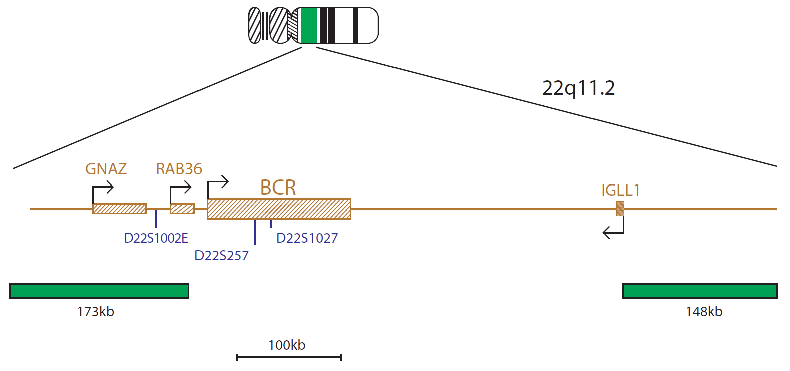
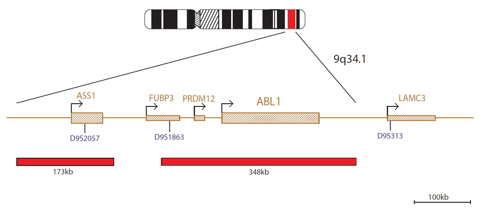
The red probe mix contains a 348kb probe that spans the ABL1 gene and a 173kb probe that spans the ASS1 gene. The green probe mix contains a 173kb probe centromeric to the BCR gene that spans the GNAZ and RAB36 genes. A second green probe covers a 148kb region telomeric to the BCR gene that spans part of the IGLL1 gene.
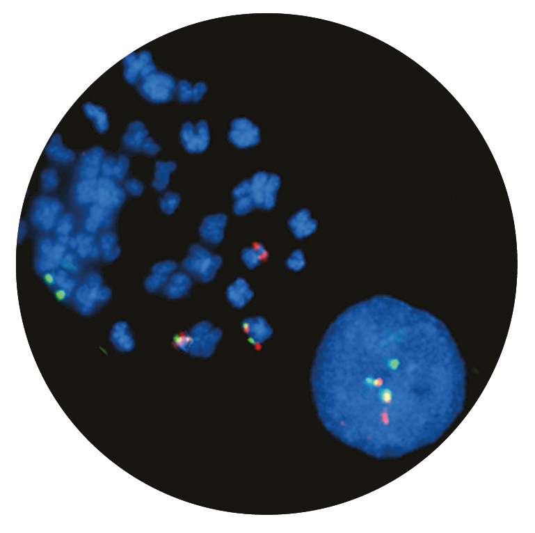
Microscope image
The BCR (BCR activator of RhoGEF and GTPase) gene is located at 22q11.2 and the ABL1 (ABL proto-oncogene 1, non-receptor tyrosine kinase) gene is located at 9q34.1. Translocation between these two genes gives rise to the BCR::ABL1 fusion gene, and produces a Philadelphia chromosome; the visible result of this translocation.
The presence of a BCR::ABL1 fusion has important diagnostic and prognostic implications in a number of haematological disorders.
The t(9;22)(q34.1;q11.2) translocation is the hallmark of chronic myeloid leukaemia (CML) and is found in around 90-95% of cases1. The remaining cases have a variant translocation, or have a cryptic rearrangement involving 9q34.1 and 22q11.2 that cannot be identified by routine cytogenetic analysis1.
The BCR::ABL1 fusion can also be found in 25% of adult acute lymphoblastic leukaemia (ALL) and in 2-4% of childhood ALL1. The presence of a BCR::ABL1 fusion has been shown to confer a poor prognosis in ALL in both adults and children1,2. The detection of the abnormality is therefore of high importance for risk stratification, which will influence treatment and management decisions2. In a small number of ALL cases, the translocation does not result in a cytogenetically visible Philadelphia chromosome. In these cases, FISH is essential for highlighting the fusion gene3.
This rearrangement is also seen in rare cases of acute myeloid leukaemia (AML). Philadelphia-positive AML is characterised by its resistance to conventional standard chemotherapy and poor prognosis4, so accurate and rapid identification of this chromosomal abnormality is vital.
The CytoCell® BCR/ABL (ABL1) Translocation, Dual Fusion Probe is a qualitative, non-automated, fluorescence in situ hybridisation (FISH) test used to detect chromosomal rearrangements between the 22q11.2 region on chromosome 22 and the 9q34.1 region on chromosome 9 in Carnoy’s solution (3:1 methanol/acetic acid) fixed haematologically-derived cell suspensions from patients with confirmed or suspected chronic myeloid leukaemia (CML), acute myeloid leukaemia (AML) or acute lymphoblastic leukaemia (ALL).
This device is designed as an adjunct to other clinical and histopathological tests in recognised diagnostic and clinical care pathways, where knowledge of BCR::ABL1 translocation status would be important for clinical management.
This device is designed to detect rearrangements with breakpoints in the region covered by the red and green clones in this probe set, which includes the BCR and ABL1 regions. Breakpoints outside this region, or variant rearrangements wholly contained within this region, may not be detected with this device.
This device is not intended for: use as a stand-alone diagnostic, use as a companion diagnostic, prenatal testing, population-based screening, near-patient testing, or self-testing.
This device has not been validated for sample types, disease types, or purposes outside of those stated in the intended purpose.
It is intended as an adjunct to other diagnostic laboratory tests and therapeutic action should not be initiated on the basis of the FISH result alone.
Reporting and interpretation of FISH results should be performed by suitably qualified staff, consistent with professional standards of practice, and should take into consideration other relevant test results, clinical and diagnostic information.
This device is intended for laboratory professional use only.
Failure to adhere to the protocol may affect the performance and lead to false positive/negative results.
Find certificate of analysis documentation for our CytoCell FISH probes

Our lab has been using a wide range of CytoCell FISH probes for a number of years, and have been increasing this range all the time. The probes have clear bright signals and show good reproducibility. CytoCell provides fast delivery of catalogue probes, and are very responsive when we have any queries or problems with their products.

Bridget Manasse
Addenbrookes Hospital, Cambridge University Hosiptals NHS Foundation Trust, UK
In our hands, CytoCell FISH probes have proven to be of the highest quality with bright, easy to interpret signals, thus providing confidence in our results. OGT's customer support is outstanding, as their staff are extremely knowledgeable and truly care about their customers and their customers’ needs.

Jennie Thurston
Director of Cytogenetics, Carolinas Pathology Group, USA
I first came across CytoCell FISH probes in a previous lab I worked in and I was struck by the quality of the products. Since this time, I have been recommending and introducing CytoCell probes across all application areas — now they are the primary FISH probes used in our lab. They have an excellent range of products and their ready-to-use reagent format saves considerable time.

Elizabeth Benner
Medical Technologist, University of Arizona Health Network, USA
We have been working with CytoCell fish probes for two decades because of their excellent clarity and intensity regardless of the size of the probe. It is so clear and simple to detect.
Dr. Marina Djurisic
Head of Laboratory of Medical Genetics, Mother and Child Health Care Institute of Serbia “Dr Vukan Cupic”, Serbia
The quality and consistency of CytoCell’s probes means I can trust the results, and my clients get their results in a timely manner.

Dr. Theresa C. Brown
Director, Cytogenetics Laboratory, Hayward Genetics Center, Tulane University School of Medicine, USA
It was very important for us to have more consistent results with our probes — easy-to-read bright signals and a range of vial sizes, which is much more cost-effective.

Janet Cowan, PhD
Director of the Cytogenetics Laboratory, Tufts Medical Center, USA
Not only do CytoCell offer an extensive range of high-quality FISH probes, the customer support is also excellent — providing fast access to all the probes I need. The probes are highly consistent with bright signals allowing easy scoring of results.
Dr. Eric Crawford
Senior Director, Genetics Associates Inc., USA
The quality and reproducibility of results using the CytoCell kit has been vital in accurately detecting co-deletions in our glioma investigations. We now have a cost-effective test that we can rely on that is also easy to use and interpret. We've been consistently impressed with this kit - not to mention the support offered by OGT's customer service, and have completely transitioned over to CytoCell probes.
Gavin Cuthbert, FRCPath
Head of Cancer Cytogenetics, Northern Genetics Servce, Newcastle, UK
 Visit USA site
 Visit Canada site


