


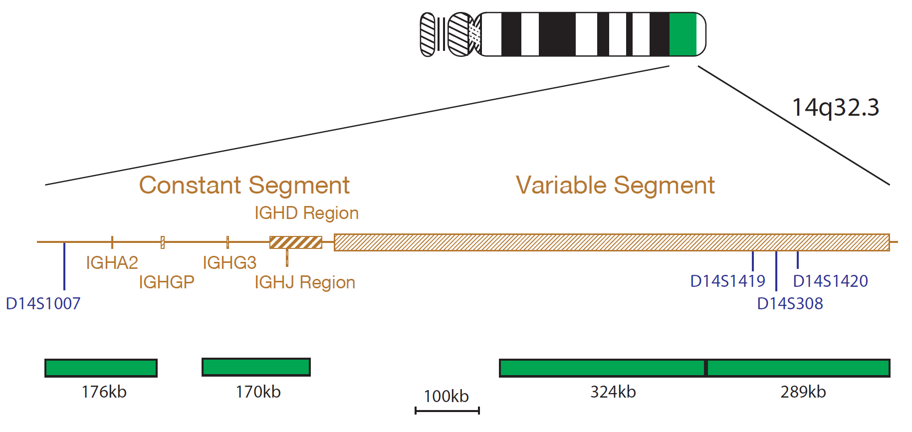
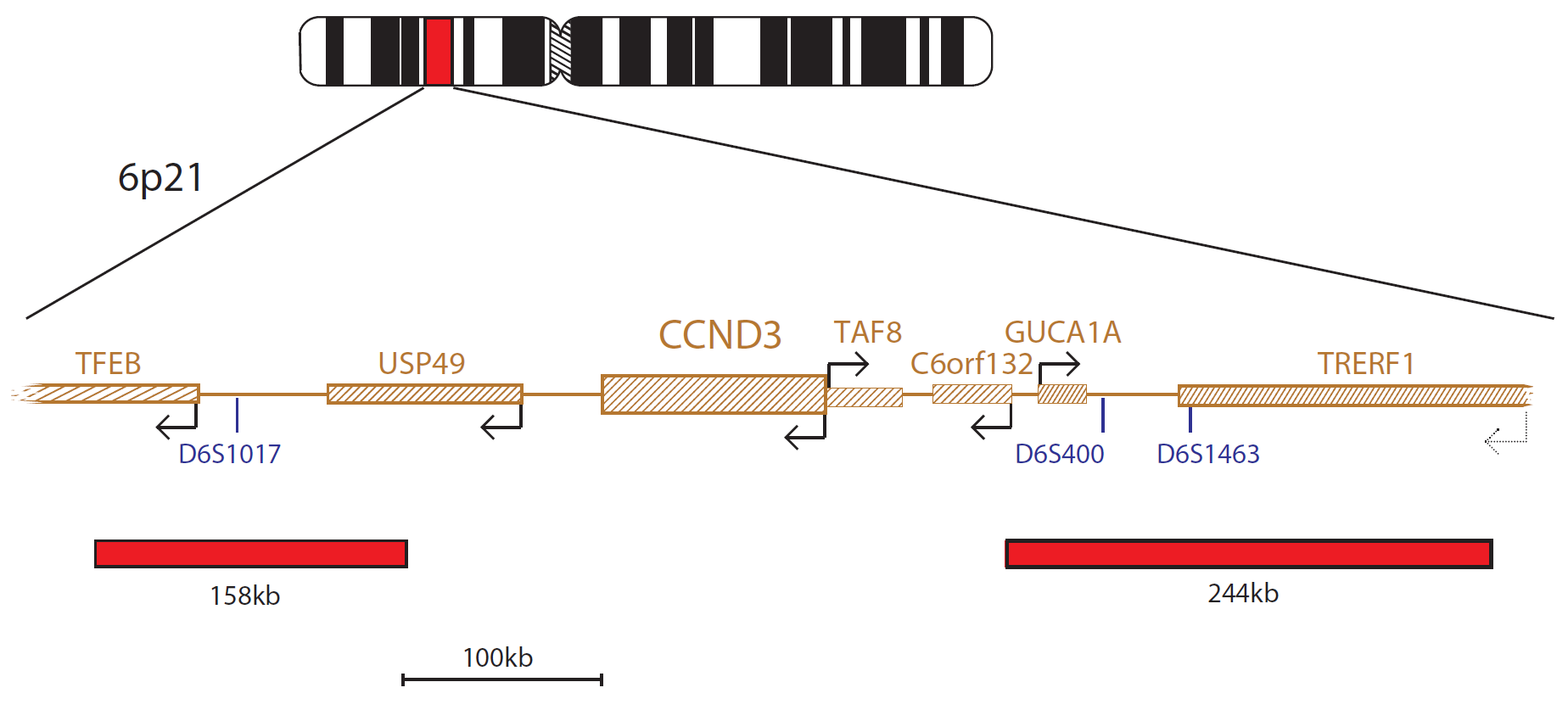
The IGH/CCND3 product consists of probes labelled in green, covering the Constant, J, D and Variable segments of the IGH gene, and CCND3 probes, labelled in red. The CCND3 probe mix contains a 158kb probe telomeric to the CCND3 gene, including the D6S1017 marker and a second probe covering the 244kb region centromeric to the TAF8 gene, including the D6S400 and the D6S1463 markers.
The CCND3 (cyclin D3) gene is located at 6p21.1 and IGH (immunoglobulin heavy locus) at 14q32.33.
Approximately 50-60% of multiple myeloma (MM) cases are associated with translocations involving IGH and one of several partners including CCND1, NSD2 (MMSET) and FGFR3, CCND3, MAF or MAFB1.
The t(6;14)(p21;q32) translocation is a recurrent translocation seen in 4% of cases of MM2.
CCND3 has been identified as a putative oncogene that is dysregulated as a consequence of the t(6;14)(p21;q32) translocation2. The translocation appears to be mediated by an error in IgH switch recombination as it has been shown that in KMM-1 cell lines, the translocation disrupts a switch sequence in this region and results in juxtaposition of CCND3 with the IGH promoter, thus elevating the levels of CCND3 expression2. It is thought that this mechanism is similar in all cases of IGH translocation. Most breakpoints appear to be clustered in a region that is fewer than 200kb centromeric to CCND32.
CCND3-IGH translocations are also reported in a variety of other B-cell malignancies, including plasma cell leukaemia, diffuse large B-cell lymphoma (DLBCL) and splenic lymphomas with villous lymphocytes (SLVL)3.
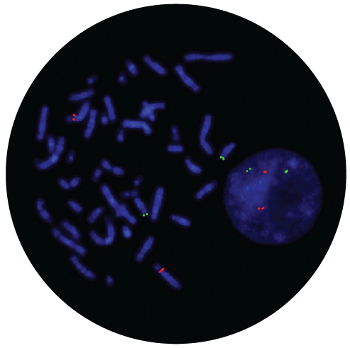
Microscope image
Find certificate of analysis documentation for our CytoCell FISH probes

Our lab has been using a wide range of CytoCell FISH probes for a number of years, and have been increasing this range all the time. The probes have clear bright signals and show good reproducibility. CytoCell provides fast delivery of catalogue probes, and are very responsive when we have any queries or problems with their products.

Bridget Manasse
Addenbrookes Hospital, Cambridge University Hosiptals NHS Foundation Trust, UK
In our hands, CytoCell FISH probes have proven to be of the highest quality with bright, easy to interpret signals, thus providing confidence in our results. OGT's customer support is outstanding, as their staff are extremely knowledgeable and truly care about their customers and their customers’ needs.

Jennie Thurston
Director of Cytogenetics, Carolinas Pathology Group, USA
I first came across CytoCell FISH probes in a previous lab I worked in and I was struck by the quality of the products. Since this time, I have been recommending and introducing CytoCell probes across all application areas — now they are the primary FISH probes used in our lab. They have an excellent range of products and their ready-to-use reagent format saves considerable time.

Elizabeth Benner
Medical Technologist, University of Arizona Health Network, USA
We have been working with CytoCell fish probes for two decades because of their excellent clarity and intensity regardless of the size of the probe. It is so clear and simple to detect.
Dr. Marina Djurisic
Head of Laboratory of Medical Genetics, Mother and Child Health Care Institute of Serbia “Dr Vukan Cupic”, Serbia
The quality and consistency of CytoCell’s probes means I can trust the results, and my clients get their results in a timely manner.

Dr. Theresa C. Brown
Director, Cytogenetics Laboratory, Hayward Genetics Center, Tulane University School of Medicine, USA
It was very important for us to have more consistent results with our probes — easy-to-read bright signals and a range of vial sizes, which is much more cost-effective.

Janet Cowan, PhD
Director of the Cytogenetics Laboratory, Tufts Medical Center, USA
Not only do CytoCell offer an extensive range of high-quality FISH probes, the customer support is also excellent — providing fast access to all the probes I need. The probes are highly consistent with bright signals allowing easy scoring of results.
Dr. Eric Crawford
Senior Director, Genetics Associates Inc., USA
The quality and reproducibility of results using the CytoCell kit has been vital in accurately detecting co-deletions in our glioma investigations. We now have a cost-effective test that we can rely on that is also easy to use and interpret. We've been consistently impressed with this kit - not to mention the support offered by OGT's customer service, and have completely transitioned over to CytoCell probes.
Gavin Cuthbert, FRCPath
Head of Cancer Cytogenetics, Northern Genetics Servce, Newcastle, UK
 Visit USA site
 Visit Canada site


