


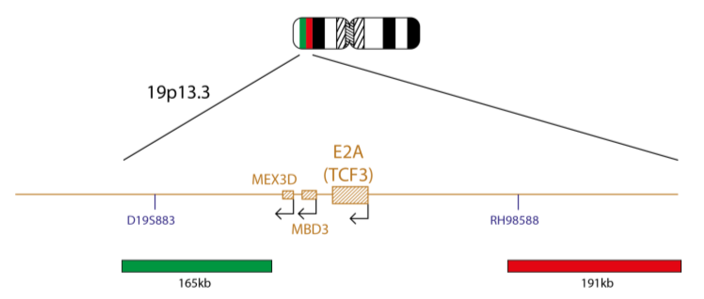
The E2A product consists of a 191kb probe, labelled in red, located centromeric to the E2A (TCF3) gene, including the RH98588 marker, and a green probe covering a 165kb region located telomeric to the E2A gene, including the D19S883 marker.
The TCF3 (transcription factor 3) gene is located at 19p13.3. Translocations involving TCF3 are some of the most common rearrangements in childhood B-cell acute lymphoblastic leukaemia (ALL)1,2.
Two of the main TCF3 partners are PBX1 (PBX homeobox 1) at 1q23.3 and HLF (HLF transcription factor, PAR bZIP family member) at 17q22. These become fused to TCF3 as a result of the t(1;19)(q23;p13) and t(17;19)(q22;p13) translocations, forming the TCF3-PBX1 and TCF3- HLF fusion genes, respectively. A rare cryptic inversion, inv(19)(p13;q13), has been reported to fuse TCF3 to TFPT (TCF3 fusion partner) resulting in the TCF3-TFPT fusion gene1.
The t(1;19)(q23;p13) is the most common TCF3 rearrangement, being present in around 6% of childhood B-ALL1,2. According to the World Health Organization (WHO) classification of myeloid neoplasms and acute leukaemia, B lymphoblastic leukaemia/lymphoma with t(1;19)(q23;p13); TCF3-PBX1 is recognised as a distinct disease entity2. The functional fusion gene resides at chromosome 19. An unbalanced form of this translocation has been reported, with loss of der(1)1,2. Detection of the E2A-PBX1 fusion by molecular methods, such as FISH, is important, as a subset of B-ALLs has a karyotypically identical t(1;19) that involves neither TCF3 nor PBX1. E2A-PBX1 positive leukaemia was historically associated with a poor outcome, though modern intensive therapies have overcome this1,2,4.
The t(17;19)(q22;p13.) is a rare translocation that is present in around 1% of precursor B-ALL cases1. TCF3-HLF positive leukaemia is associated with adverse prognosis3,4.
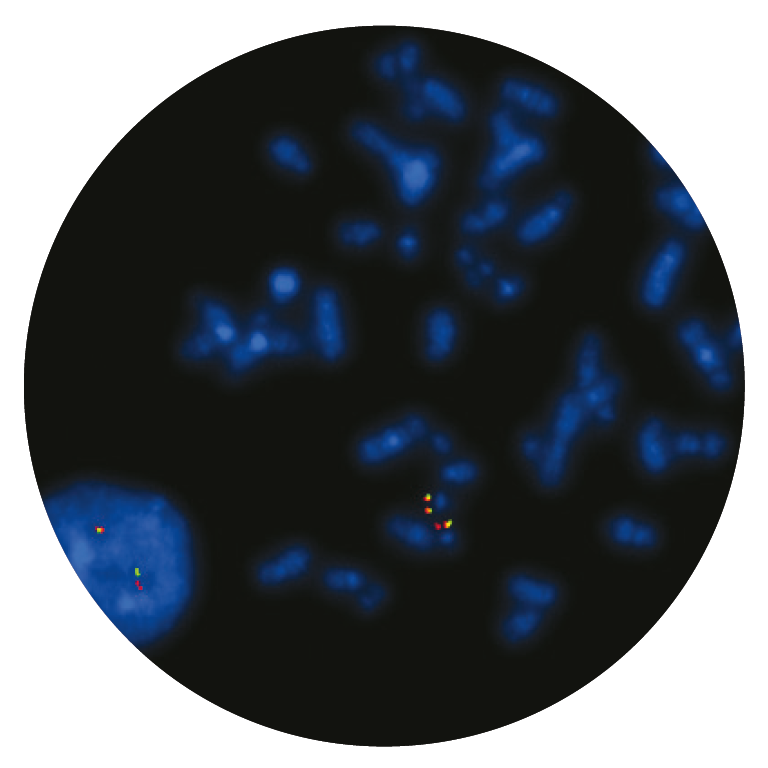
Microscope image
Find certificate of analysis documentation for our CytoCell FISH probes

Our lab has been using a wide range of CytoCell FISH probes for a number of years, and have been increasing this range all the time. The probes have clear bright signals and show good reproducibility. CytoCell provides fast delivery of catalogue probes, and are very responsive when we have any queries or problems with their products.

Bridget Manasse
Addenbrookes Hospital, Cambridge University Hosiptals NHS Foundation Trust, UK
In our hands, CytoCell FISH probes have proven to be of the highest quality with bright, easy to interpret signals, thus providing confidence in our results. OGT's customer support is outstanding, as their staff are extremely knowledgeable and truly care about their customers and their customers’ needs.

Jennie Thurston
Director of Cytogenetics, Carolinas Pathology Group, USA
I first came across CytoCell FISH probes in a previous lab I worked in and I was struck by the quality of the products. Since this time, I have been recommending and introducing CytoCell probes across all application areas — now they are the primary FISH probes used in our lab. They have an excellent range of products and their ready-to-use reagent format saves considerable time.

Elizabeth Benner
Medical Technologist, University of Arizona Health Network, USA
We have been working with CytoCell fish probes for two decades because of their excellent clarity and intensity regardless of the size of the probe. It is so clear and simple to detect.
Dr. Marina Djurisic
Head of Laboratory of Medical Genetics, Mother and Child Health Care Institute of Serbia “Dr Vukan Cupic”, Serbia
The quality and consistency of CytoCell’s probes means I can trust the results, and my clients get their results in a timely manner.

Dr. Theresa C. Brown
Director, Cytogenetics Laboratory, Hayward Genetics Center, Tulane University School of Medicine, USA
It was very important for us to have more consistent results with our probes — easy-to-read bright signals and a range of vial sizes, which is much more cost-effective.

Janet Cowan, PhD
Director of the Cytogenetics Laboratory, Tufts Medical Center, USA
Not only do CytoCell offer an extensive range of high-quality FISH probes, the customer support is also excellent — providing fast access to all the probes I need. The probes are highly consistent with bright signals allowing easy scoring of results.
Dr. Eric Crawford
Senior Director, Genetics Associates Inc., USA
The quality and reproducibility of results using the CytoCell kit has been vital in accurately detecting co-deletions in our glioma investigations. We now have a cost-effective test that we can rely on that is also easy to use and interpret. We've been consistently impressed with this kit - not to mention the support offered by OGT's customer service, and have completely transitioned over to CytoCell probes.
Gavin Cuthbert, FRCPath
Head of Cancer Cytogenetics, Northern Genetics Servce, Newcastle, UK
 Visit USA site
 Visit Canada site


