The study of chromosomes in the detection of genetic abnormalities is called cytogenetics. Fluorescence in situ hybridization (FISH) combines conventional cytogenetics with molecular genetics and can be used in hematology, pathology and constitutional cytogenetics.
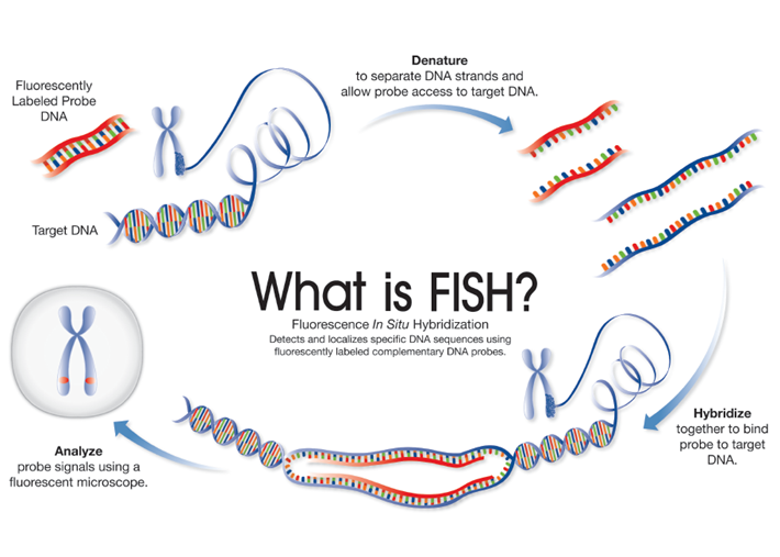
FISH is a technique that uses fluorescently labelled DNA sequences (probes) to assess specific regions of interest, such as genes and centromeres using fluorescent microscopy.
The DNA sequence of the FISH probe is complementary to the region of interest. When using a FISH probe, if the region of interest is present, the probe will bind (hybridize) to the target DNA sequence, which can then be visualized using fluorescent microscopy – indicating the presence of the gene/centromere etc. If the region of interest has been deleted, the probe will not bind to the target DNA and so will not be visualized. A breakapart or a translocation FISH probe can be used to detect whether a gene has been rearranged.
