


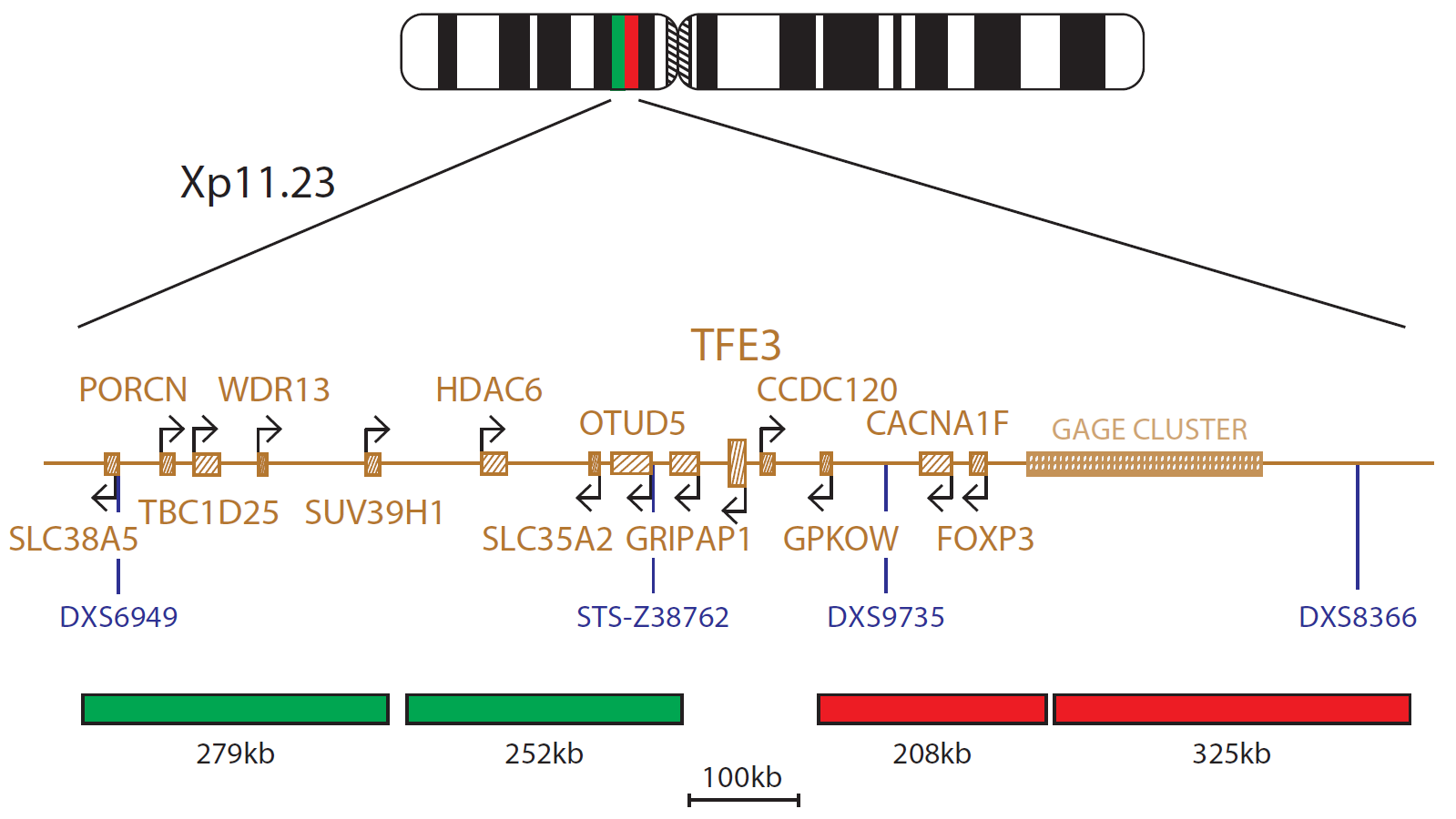
The TFE3 Breakapart probe consists of two probes (279kb and 252kb), labelled in green, situated distal to the TFE3 gene and covering markers DXS6949 and STS-Z38762 and two probes (208kb and 325kb), labelled in red, situated proximal to the TFE3 gene and covering markers DXS9735 and DXS8366.
TFE3 (transcription factor binding to IGHM enhancer 3) is a protein-coding gene located at Xp11.23. Recurrent rearrangements of the TFE3 gene have been reported in a number of neoplastic diseases – often grouped together as ‘Xp11 translocation cancers’ – these include: renal cell carcinoma (RCC), soft tissue alveolar soft part sarcoma (ASPS), perivascular epithelioid cell tumors (PEComa), epithelioid hemangioendotheliomas (EHE) and melanotic Xp11 translocation renal cancer1,2.
In RCC, TFE3 translocation partners have been shown to include the ASPSCR1, SFPQ and NONO genes3. Although RCC and ASPS have been shown to have identical ASPSCR1-TFE3 fusion transcripts, the t(X;17) translocation is consistently balanced in the former but usually unbalanced in the latter - the derivative X chromosome is not seen in ASPS4.
In epithelioid hemangioendothelioma the novel YAP1-TFE3 fusion is seen, and defines a clinically distinct subset of this disease5,6, whereas in PEComa, the predominant partner has been shown to be the SFPQ gene7.
This research use only (RUO) probe has been designed for the investigation of TFE3 rearrangements, regardless of the partner gene involved.
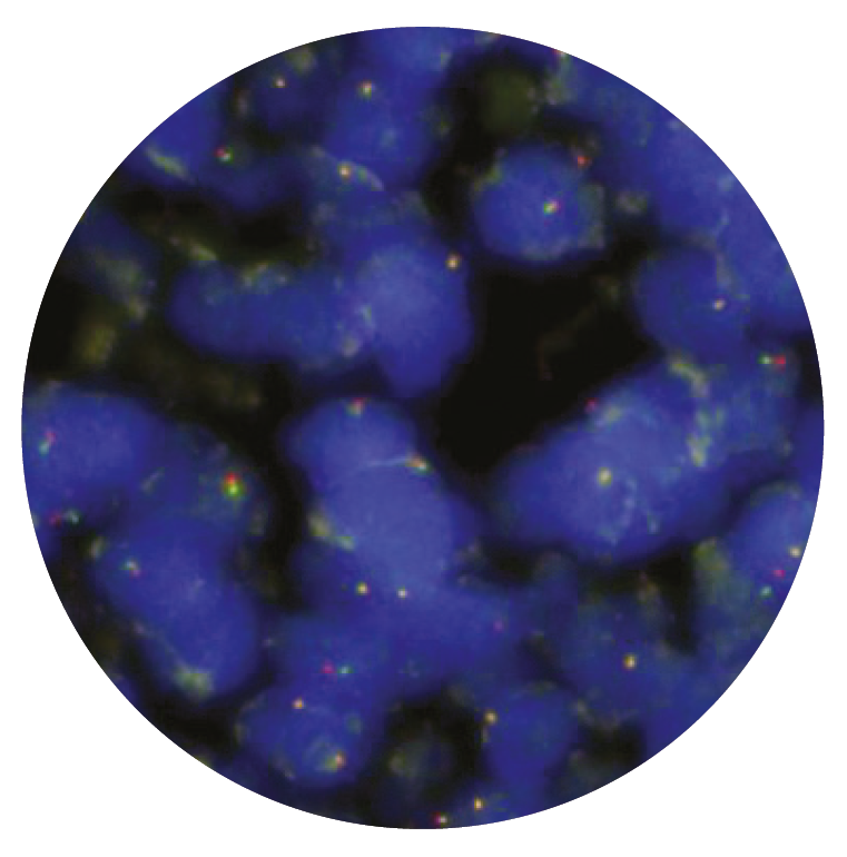
Microscope image
Research use only (RUO)
Find certificate of analysis documentation for our CytoCell FISH probes

Our lab has been using a wide range of CytoCell FISH probes for a number of years, and have been increasing this range all the time. The probes have clear bright signals and show good reproducibility. CytoCell provides fast delivery of catalogue probes, and are very responsive when we have any queries or problems with their products.

Bridget Manasse
Addenbrookes Hospital, Cambridge University Hosiptals NHS Foundation Trust, UK
In our hands, CytoCell FISH probes have proven to be of the highest quality with bright, easy to interpret signals, thus providing confidence in our results. OGT's customer support is outstanding, as their staff are extremely knowledgeable and truly care about their customers and their customers’ needs.

Jennie Thurston
Director of Cytogenetics, Carolinas Pathology Group, USA
I first came across CytoCell FISH probes in a previous lab I worked in and I was struck by the quality of the products. Since this time, I have been recommending and introducing CytoCell probes across all application areas — now they are the primary FISH probes used in our lab. They have an excellent range of products and their ready-to-use reagent format saves considerable time.

Elizabeth Benner
Medical Technologist, University of Arizona Health Network, USA
We have been working with CytoCell fish probes for two decades because of their excellent clarity and intensity regardless of the size of the probe. It is so clear and simple to detect.
Dr. Marina Djurisic
Head of Laboratory of Medical Genetics, Mother and Child Health Care Institute of Serbia “Dr Vukan Cupic”, Serbia
The quality and consistency of CytoCell’s probes means I can trust the results, and my clients get their results in a timely manner.

Dr. Theresa C. Brown
Director, Cytogenetics Laboratory, Hayward Genetics Center, Tulane University School of Medicine, USA
It was very important for us to have more consistent results with our probes — easy-to-read bright signals and a range of vial sizes, which is much more cost-effective.

Janet Cowan, PhD
Director of the Cytogenetics Laboratory, Tufts Medical Center, USA
Not only do CytoCell offer an extensive range of high-quality FISH probes, the customer support is also excellent — providing fast access to all the probes I need. The probes are highly consistent with bright signals allowing easy scoring of results.
Dr. Eric Crawford
Senior Director, Genetics Associates Inc., USA
The quality and reproducibility of results using the CytoCell kit has been vital in accurately detecting co-deletions in our glioma investigations. We now have a cost-effective test that we can rely on that is also easy to use and interpret. We've been consistently impressed with this kit - not to mention the support offered by OGT's customer service, and have completely transitioned over to CytoCell probes.
Gavin Cuthbert, FRCPath
Head of Cancer Cytogenetics, Northern Genetics Servce, Newcastle, UK
 Visit USA site
 Visit Canada site


