


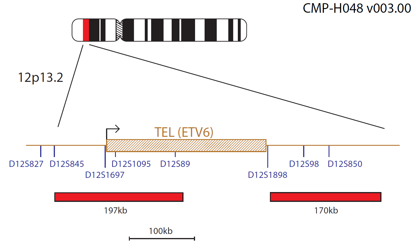
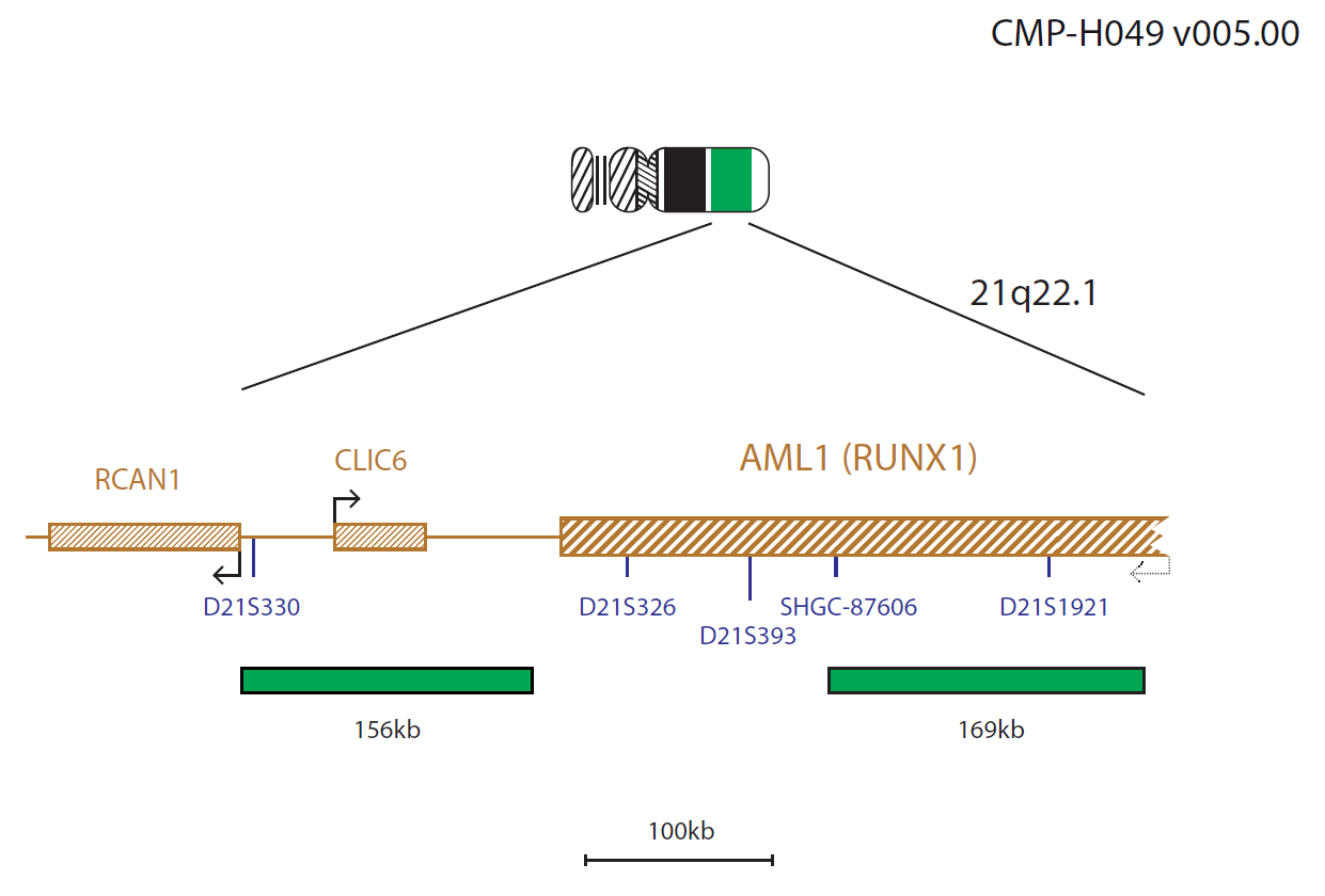
The TEL probe mix, labelled in red, contains a probe covering a 197kb region at the telomeric end of TEL (ETV6) and a second probe that extends 170kb in a centromeric direction beyond the TEL (ETV6) gene. The AML1 (RUNX1) probe set consists of two probes labelled in green. One locates to a 156kb region centromeric to the AML1 (RUNX1) gene and covers the CLIC6 gene. The other probe covers a 169kb region in the AML1 (RUNX1) gene that includes the markers SHGC-87606 and D21S1921.
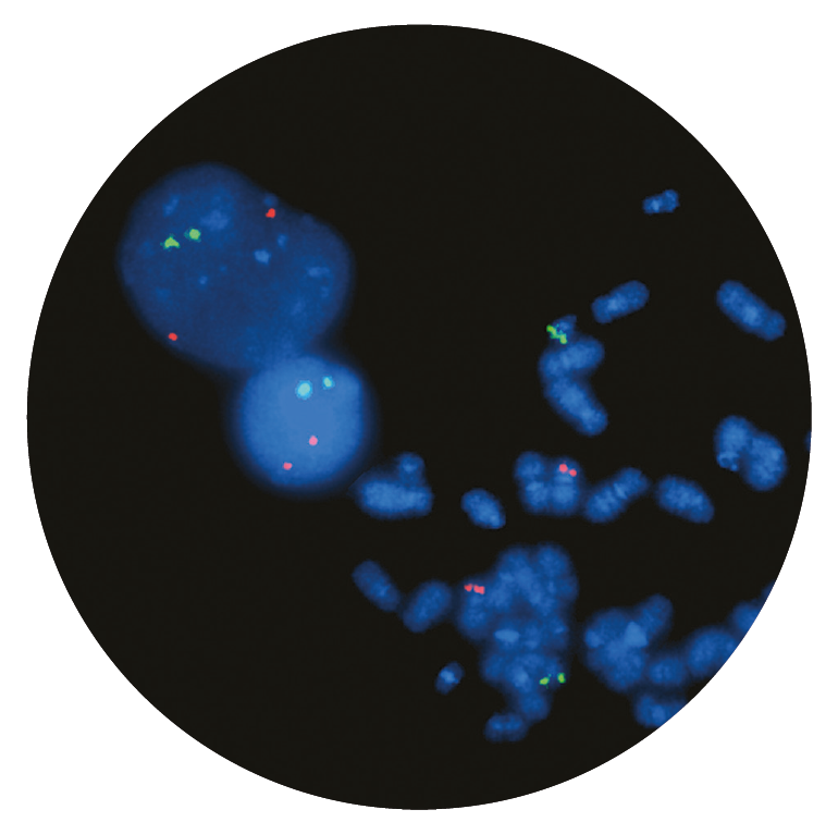
The cytogenetically-cryptic t(12;21)(p13;q22) translocation between ETV6 (ets variant 6) at 12p13 and RUNX1, (RUNX family transcription factor 1) at 21q22, results in the ETV6-RUNX1 chimeric fusion gene1.
The ETV6 and RUNX1 genes both encode transcription factors; ETV6 has been shown to be required for proper transcription during haematopoiesis within the bone marrow1,2. The ETV6-RUNX1 protein converts RUNX1 to a transcriptional repressor and causes overexpression of the erythropoietin receptor (EPOR) and activation of downstream JAK-STAT signaling1.
B-lymphoblastic leukaemia/lymphomas with t(12;21)(p13;q22) translocations form a recognised disease entity according to the World Health Organization (WHO) classification of myeloid neoplasms and acute leukaemia. This is the most common sub-group of childhood B-ALL accounting for about 25% of cases3. As the t(12;21)(p13;q22) translocation is cytogenetically-cryptic, FISH is an important diagnostic tool for this leukaemia4.
B-ALL with ETV6-RUNX1 is considered to have a favourable outcome with cure rates more than 90%3. Late relapses have been reported; these have been attributed to the presence of persistent preleukaemic clones that survived chemotherapy3,5.
ETV6 has also been shown to be deleted in some children with ALL, with loss of heterozygosity (LOH) of chromosome 12p12-13; these deletions often seen in the presence of ETV6-RUNX1 translocations6.
Microscope image
Find certificate of analysis documentation for our CytoCell FISH probes

Our lab has been using a wide range of CytoCell FISH probes for a number of years, and have been increasing this range all the time. The probes have clear bright signals and show good reproducibility. CytoCell provides fast delivery of catalogue probes, and are very responsive when we have any queries or problems with their products.

Bridget Manasse
Addenbrookes Hospital, Cambridge University Hosiptals NHS Foundation Trust, UK
In our hands, CytoCell FISH probes have proven to be of the highest quality with bright, easy to interpret signals, thus providing confidence in our results. OGT's customer support is outstanding, as their staff are extremely knowledgeable and truly care about their customers and their customers’ needs.

Jennie Thurston
Director of Cytogenetics, Carolinas Pathology Group, USA
I first came across CytoCell FISH probes in a previous lab I worked in and I was struck by the quality of the products. Since this time, I have been recommending and introducing CytoCell probes across all application areas — now they are the primary FISH probes used in our lab. They have an excellent range of products and their ready-to-use reagent format saves considerable time.

Elizabeth Benner
Medical Technologist, University of Arizona Health Network, USA
We have been working with CytoCell fish probes for two decades because of their excellent clarity and intensity regardless of the size of the probe. It is so clear and simple to detect.
Dr. Marina Djurisic
Head of Laboratory of Medical Genetics, Mother and Child Health Care Institute of Serbia “Dr Vukan Cupic”, Serbia
The quality and consistency of CytoCell’s probes means I can trust the results, and my clients get their results in a timely manner.

Dr. Theresa C. Brown
Director, Cytogenetics Laboratory, Hayward Genetics Center, Tulane University School of Medicine, USA
It was very important for us to have more consistent results with our probes — easy-to-read bright signals and a range of vial sizes, which is much more cost-effective.

Janet Cowan, PhD
Director of the Cytogenetics Laboratory, Tufts Medical Center, USA
Not only do CytoCell offer an extensive range of high-quality FISH probes, the customer support is also excellent — providing fast access to all the probes I need. The probes are highly consistent with bright signals allowing easy scoring of results.
Dr. Eric Crawford
Senior Director, Genetics Associates Inc., USA
The quality and reproducibility of results using the CytoCell kit has been vital in accurately detecting co-deletions in our glioma investigations. We now have a cost-effective test that we can rely on that is also easy to use and interpret. We've been consistently impressed with this kit - not to mention the support offered by OGT's customer service, and have completely transitioned over to CytoCell probes.
Gavin Cuthbert, FRCPath
Head of Cancer Cytogenetics, Northern Genetics Servce, Newcastle, UK
 Visit USA site
 Visit Canada site


