


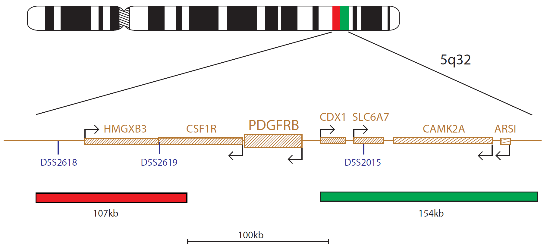
The PDGFRB product consists of a 107kb probe, labelled in red, located centromeric to the PDGFRB gene, including the D5S2618 and D5S2619 markers and a green probe, covering a 154kb region telomeric to the PDGFRB gene, including the D5S2015 marker.
Rearrangements involving the PDGFRB (platelet derived growth factor receptor beta) gene at 5q32 are reported in both myeloid and lymphoid neoplasms.
In the 2008 World Health Organization (WHO) classification of myeloid neoplasms and acute leukaemia, a new subgroup of myeloid neoplasms was introduced: Myeloid and Lymphoid Neoplasms with Eosinophilia and Abnormalities of PDGFRA, PDGFRB or FGFR1. These neoplasms constitute three specific disease groups, with some shared features1.
The myeloid neoplasms with PDGFRB rearrangements are characterised by constitutive activation of the PDGFRB gene product1. The activation is most commonly caused by a t(5;12)(q31-q33;p13) translocation which results in an ETV6-PDGFRB fusion gene. Patients with this fusion have been shown to be responsive to tyrosine kinase inhibitors (TKIs)2, which specifically inhibits the kinase activity of PDGFRB.
In B-ALL, gene expression profiling has identified an unusual genetic subgroup, the BCR-ABL1-like or Philadelphia chromosome-like (Ph-like) ALL, which represents about 15% of paediatric ALL cases and has an unfavourable outcome3,4. Patients with this expression signature are characterised by genetic alterations, such as rearrangements, mutations and deletions of a range of kinase and cytokine receptors, including PDGFRB rearrangements. Known PDGFRB partners are EBF1 at 5q33, SSBP2 at 5q14, TNIP1 at 5q33 and ZEB2 at 2q22. It is crucial to detect such rearrangements, as patients could benefit from treatment with tyrosine kinase inhibitors (TKIs)3,4,5.
Microscope image
Find certificate of analysis documentation for our CytoCell FISH probes

Our lab has been using a wide range of CytoCell FISH probes for a number of years, and have been increasing this range all the time. The probes have clear bright signals and show good reproducibility. CytoCell provides fast delivery of catalogue probes, and are very responsive when we have any queries or problems with their products.

Bridget Manasse
Addenbrookes Hospital, Cambridge University Hosiptals NHS Foundation Trust, UK
In our hands, CytoCell FISH probes have proven to be of the highest quality with bright, easy to interpret signals, thus providing confidence in our results. OGT's customer support is outstanding, as their staff are extremely knowledgeable and truly care about their customers and their customers’ needs.

Jennie Thurston
Director of Cytogenetics, Carolinas Pathology Group, USA
I first came across CytoCell FISH probes in a previous lab I worked in and I was struck by the quality of the products. Since this time, I have been recommending and introducing CytoCell probes across all application areas — now they are the primary FISH probes used in our lab. They have an excellent range of products and their ready-to-use reagent format saves considerable time.

Elizabeth Benner
Medical Technologist, University of Arizona Health Network, USA
We have been working with CytoCell fish probes for two decades because of their excellent clarity and intensity regardless of the size of the probe. It is so clear and simple to detect.
Dr. Marina Djurisic
Head of Laboratory of Medical Genetics, Mother and Child Health Care Institute of Serbia “Dr Vukan Cupic”, Serbia
The quality and consistency of CytoCell’s probes means I can trust the results, and my clients get their results in a timely manner.

Dr. Theresa C. Brown
Director, Cytogenetics Laboratory, Hayward Genetics Center, Tulane University School of Medicine, USA
It was very important for us to have more consistent results with our probes — easy-to-read bright signals and a range of vial sizes, which is much more cost-effective.

Janet Cowan, PhD
Director of the Cytogenetics Laboratory, Tufts Medical Center, USA
Not only do CytoCell offer an extensive range of high-quality FISH probes, the customer support is also excellent — providing fast access to all the probes I need. The probes are highly consistent with bright signals allowing easy scoring of results.
Dr. Eric Crawford
Senior Director, Genetics Associates Inc., USA
The quality and reproducibility of results using the CytoCell kit has been vital in accurately detecting co-deletions in our glioma investigations. We now have a cost-effective test that we can rely on that is also easy to use and interpret. We've been consistently impressed with this kit - not to mention the support offered by OGT's customer service, and have completely transitioned over to CytoCell probes.
Gavin Cuthbert, FRCPath
Head of Cancer Cytogenetics, Northern Genetics Servce, Newcastle, UK
 Visit USA site
 Visit Canada site


