


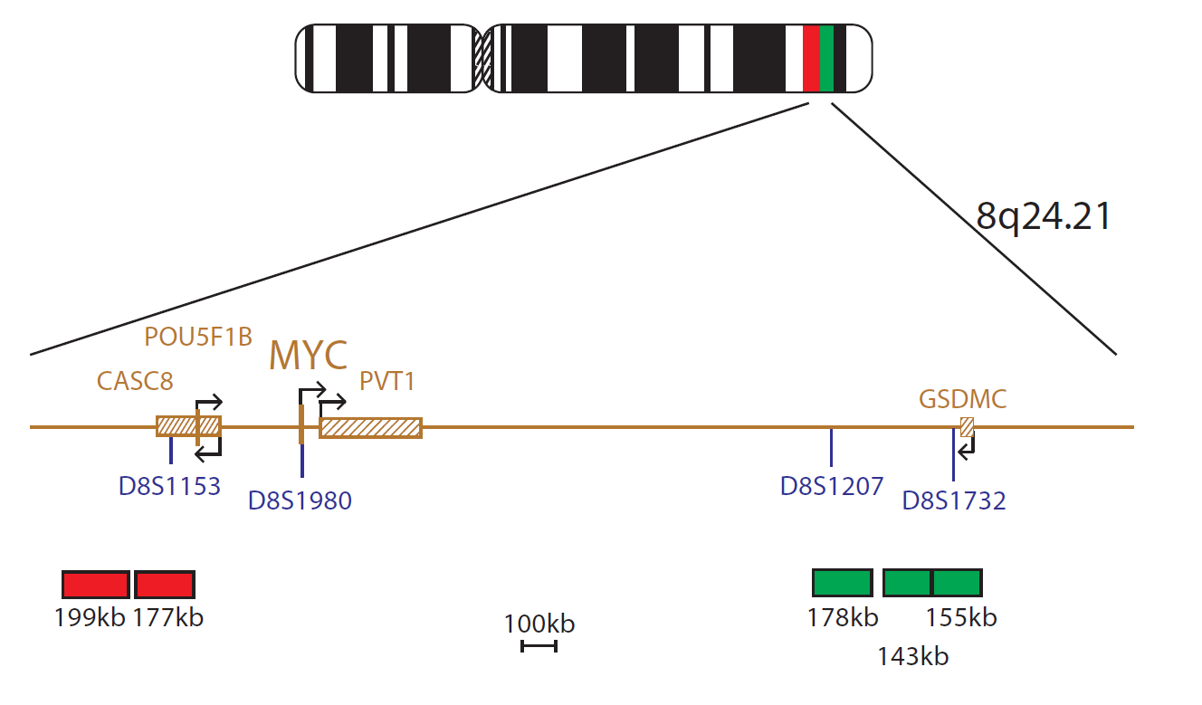
This probe set consists of two red probes that are centromeric to the MYC gene and cover the marker D8S1153 and three green probes that are telomeric to the MYC gene and cover the markers D8S1207 and D8S1732.
Translocations involving the MYC (cMYC) oncogene (Avian Myelocytomatosis Viral Oncogene Homologue) are a molecular feature of Burkitt lymphoma and occur in almost every case. These rearrangements can also be found in 5 to 10% of diffuse large B-cell lymphoma (DLBCL)1.
The majority of rearrangements result in the juxtaposition of MYC to IGH, IGL or IGK in the t(8;14), t(8;22) or t(2;8) respectively. In each case, this results in dysregulation of MYC (as a result of being close to the constitutively active immunoglobulin locus), increased transcription and neoplastic growth2. The breakpoints involved are widely scattered throughout the gene but the t(8;14) translocation always spares the protein coding exons which become attached to the derived 14. In the t(2;8) and t(8;22), MYC remains on chromosome 8 and the immunoglobulin locus joins it, resulting in close positioning of the MYC and immunoglobulin loci. Rare cases of translocations not involving immunoglobulin partners have also been reported3.
Patients with MYC rearrangements, in isolation of a confirmed diagnosis of Burkitt lymphoma, were originally thought to have poor prognoses but they do respond well to intensive chemotherapy affording them an increased survival rate. This shows that cytogenetic confirmation of the rearrangement is necessary to manage the patient effectively4.
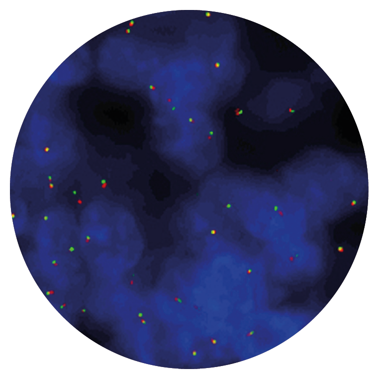
Microscope image
In vitro diagnostic (IVD)
→ English/Français/Italiano/Deutsch/Español
→ Polski
Research use only (RUO)
→ English
Find certificate of analysis documentation for our CytoCell FISH probes

Our lab has been using a wide range of CytoCell FISH probes for a number of years, and have been increasing this range all the time. The probes have clear bright signals and show good reproducibility. CytoCell provides fast delivery of catalogue probes, and are very responsive when we have any queries or problems with their products.

Bridget Manasse
Addenbrookes Hospital, Cambridge University Hosiptals NHS Foundation Trust, UK
In our hands, CytoCell FISH probes have proven to be of the highest quality with bright, easy to interpret signals, thus providing confidence in our results. OGT's customer support is outstanding, as their staff are extremely knowledgeable and truly care about their customers and their customers’ needs.

Jennie Thurston
Director of Cytogenetics, Carolinas Pathology Group, USA
I first came across CytoCell FISH probes in a previous lab I worked in and I was struck by the quality of the products. Since this time, I have been recommending and introducing CytoCell probes across all application areas — now they are the primary FISH probes used in our lab. They have an excellent range of products and their ready-to-use reagent format saves considerable time.

Elizabeth Benner
Medical Technologist, University of Arizona Health Network, USA
We have been working with CytoCell fish probes for two decades because of their excellent clarity and intensity regardless of the size of the probe. It is so clear and simple to detect.
Dr. Marina Djurisic
Head of Laboratory of Medical Genetics, Mother and Child Health Care Institute of Serbia “Dr Vukan Cupic”, Serbia
The quality and consistency of CytoCell’s probes means I can trust the results, and my clients get their results in a timely manner.

Dr. Theresa C. Brown
Director, Cytogenetics Laboratory, Hayward Genetics Center, Tulane University School of Medicine, USA
It was very important for us to have more consistent results with our probes — easy-to-read bright signals and a range of vial sizes, which is much more cost-effective.

Janet Cowan, PhD
Director of the Cytogenetics Laboratory, Tufts Medical Center, USA
Not only do CytoCell offer an extensive range of high-quality FISH probes, the customer support is also excellent — providing fast access to all the probes I need. The probes are highly consistent with bright signals allowing easy scoring of results.
Dr. Eric Crawford
Senior Director, Genetics Associates Inc., USA
The quality and reproducibility of results using the CytoCell kit has been vital in accurately detecting co-deletions in our glioma investigations. We now have a cost-effective test that we can rely on that is also easy to use and interpret. We've been consistently impressed with this kit - not to mention the support offered by OGT's customer service, and have completely transitioned over to CytoCell probes.
Gavin Cuthbert, FRCPath
Head of Cancer Cytogenetics, Northern Genetics Servce, Newcastle, UK
 Visit USA site
 Visit Canada site


