


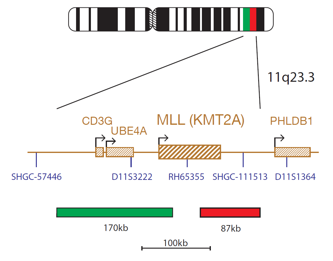
The MLL product consists of an 87kb probe, labelled in red, covering a region telomeric to the MLL (KMT2A) gene including the marker SHGC-111513 and a green probe covering a 170kb region centromeric to the MLL gene spanning the CD3G and UBE4A genes.
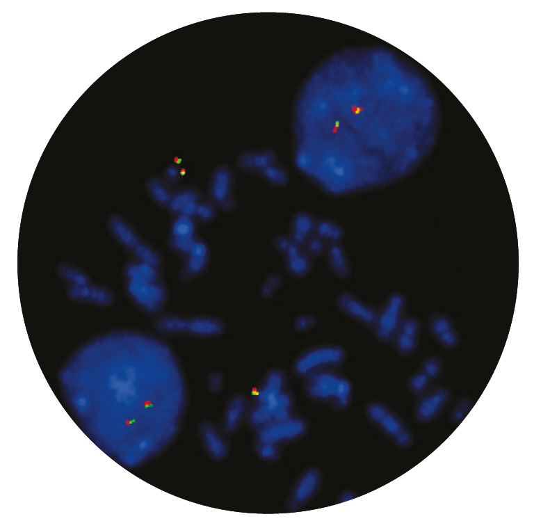
Microscope image
The KMT2A (lysine methyltransferase 2A) gene at 11q23.3 is commonly rearranged in acute leukaemias, especially in infant leukaemia and in secondary leukaemia, following treatment with DNA topoisomerase II inhibitors1.
The KMT2A gene has a great homology with the drosophila trithorax gene and encodes for a histone methyltransferase, which functions as an epigenetic regulator of transcription. KMT2A translocations result in the production of a chimeric protein in which the amino-terminal portion of KMT2A is fused to the carboxy-terminal portion of the fusion partner gene. The functional protein plays a critical role in embryonic development and haematopoiesis1,2,3,4.
KMT2A rearrangements can be detected in approximately 80% of infants with acute lymphoblastic leukaemia (ALL) and in 5-10% of paediatric and adult ALLs3,4. They can also be found in 60% of infant acute myeloid leukaemia (AML) and in 3% of de novo and 10% of therapy related adult AML cases3,5. To date, more than 70 partners have been identified with the most common translocations being MLL::AFF1; t(4;11)(q21;q23.3), MLL::MLLT4; t(6;11)(q27;q23.3), MLL::MLLT3; t(9;11) (p22;q23.3) and MLL::MLLT1; t(11;19)(q23.3;p13.3)1.
Historically, KMT2A rearrangements in acute leukaemia were associated with a poorer outcome, but recent studies have shown that the prognosis is highly dependent on the fusion partner and it may differ between children and adults1.
The CytoCell® MLL (KMT2A) Breakapart Probe is a qualitative, non-automated, fluorescence in situ hybridisation (FISH) test used to detect chromosomal rearrangements in the 11q23.3 region on chromosome 11 in Carnoy’s solution (3:1 methanol/acetic acid) fixed haematologically-derived cell suspensions from patients with confirmed or suspected acute myeloid leukaemia (AML), myelodysplastic syndromes (MDS), or acute lymphoblastic leukaemia (ALL).
This device is designed as an adjunct to other clinical and histopathological tests in recognised diagnostic and clinical care pathways, where knowledge of MLL (KMT2A) rearrangement status would be important for clinical management.
This device is designed to detect rearrangements with breakpoints in the region bounded by the red and green clones in this probe set, which includes the MLL (KMT2A) gene. Breakpoints outside of this region, or variant rearrangements wholly contained within this region, may not be detected with this device.
This device is not intended for: use as a stand-alone diagnostic, use as a companion diagnostic, prenatal testing, population-based screening, near-patient testing, or self-testing.
This device has not been validated for sample types, disease types, or purposes outside of those stated in the intended use.
It is intended as an adjunct to other diagnostic laboratory tests and therapeutic action should not be initiated on the basis of the FISH result alone.
Reporting and interpretation of FISH results should be performed by suitably qualified staff, consistent with professional standards of practice, and should take into consideration other relevant test results, clinical and diagnostic information.
This device is intended for laboratory professional use only.
Failure to adhere to the protocol may affect the performance and lead to false positive/negative results.
Find certificate of analysis documentation for our CytoCell FISH probes

Our lab has been using a wide range of CytoCell FISH probes for a number of years, and have been increasing this range all the time. The probes have clear bright signals and show good reproducibility. CytoCell provides fast delivery of catalogue probes, and are very responsive when we have any queries or problems with their products.

Bridget Manasse
Addenbrookes Hospital, Cambridge University Hosiptals NHS Foundation Trust, UK
In our hands, CytoCell FISH probes have proven to be of the highest quality with bright, easy to interpret signals, thus providing confidence in our results. OGT's customer support is outstanding, as their staff are extremely knowledgeable and truly care about their customers and their customers’ needs.

Jennie Thurston
Director of Cytogenetics, Carolinas Pathology Group, USA
I first came across CytoCell FISH probes in a previous lab I worked in and I was struck by the quality of the products. Since this time, I have been recommending and introducing CytoCell probes across all application areas — now they are the primary FISH probes used in our lab. They have an excellent range of products and their ready-to-use reagent format saves considerable time.

Elizabeth Benner
Medical Technologist, University of Arizona Health Network, USA
We have been working with CytoCell fish probes for two decades because of their excellent clarity and intensity regardless of the size of the probe. It is so clear and simple to detect.
Dr. Marina Djurisic
Head of Laboratory of Medical Genetics, Mother and Child Health Care Institute of Serbia “Dr Vukan Cupic”, Serbia
The quality and consistency of CytoCell’s probes means I can trust the results, and my clients get their results in a timely manner.

Dr. Theresa C. Brown
Director, Cytogenetics Laboratory, Hayward Genetics Center, Tulane University School of Medicine, USA
It was very important for us to have more consistent results with our probes — easy-to-read bright signals and a range of vial sizes, which is much more cost-effective.

Janet Cowan, PhD
Director of the Cytogenetics Laboratory, Tufts Medical Center, USA
Not only do CytoCell offer an extensive range of high-quality FISH probes, the customer support is also excellent — providing fast access to all the probes I need. The probes are highly consistent with bright signals allowing easy scoring of results.
Dr. Eric Crawford
Senior Director, Genetics Associates Inc., USA
The quality and reproducibility of results using the CytoCell kit has been vital in accurately detecting co-deletions in our glioma investigations. We now have a cost-effective test that we can rely on that is also easy to use and interpret. We've been consistently impressed with this kit - not to mention the support offered by OGT's customer service, and have completely transitioned over to CytoCell probes.
Gavin Cuthbert, FRCPath
Head of Cancer Cytogenetics, Northern Genetics Servce, Newcastle, UK
 Visit USA site
 Visit Canada site


