


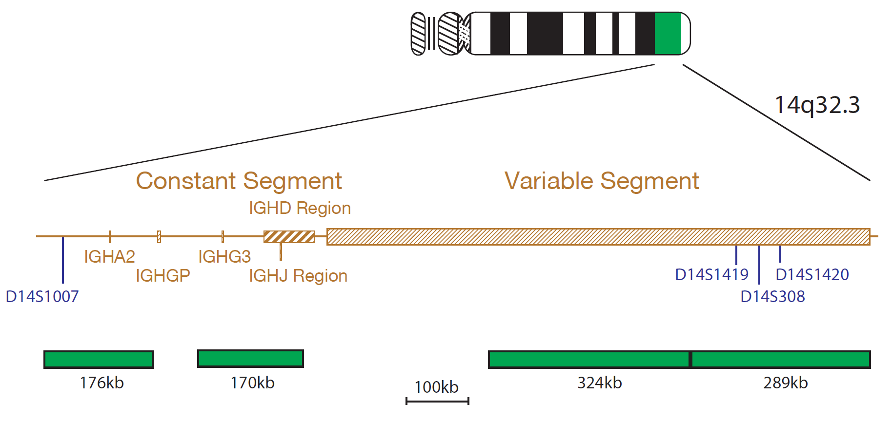
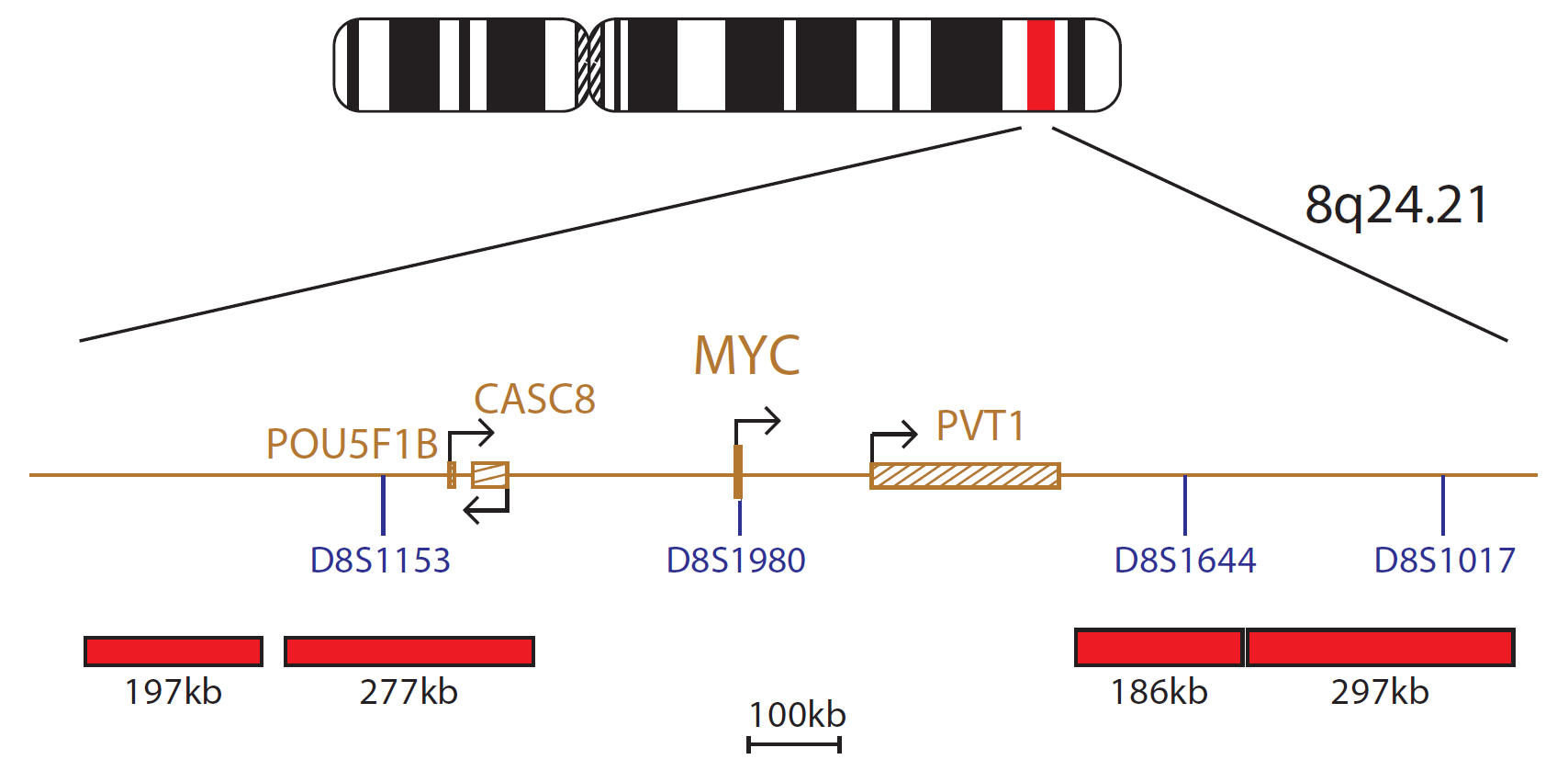
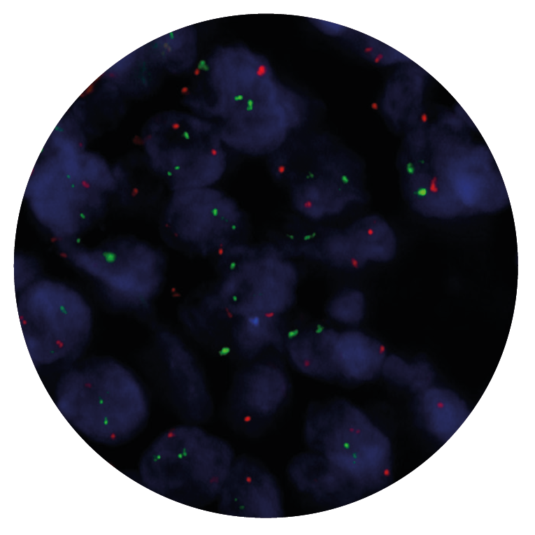
The IGH/MYC product consists of probes, labelled in green, covering the Constant, J, D and Variable segments of the IGH gene, and MYC probes, labelled in red. The MYC probe mix contains four probes, two 197kb, 277kb probes positioned centromeric to the MYC gene and two 186kb, 297kb probes positioned telomeric to the MYC gene, including the D8S1644 marker.
The cMYC/IGH translocation, t(8;14)(q24;q32), and the variant forms t(2;8)(p13;q24) and t(8;22)(q24;q11) are found in Burkitt's lymphoma and mature B-cell or Burkitt's type Acute Lymphoblastic Leukaemia (ALL)1.
The t(8;14) is the most common and is found in approximately 85% of patients with Burkitt's lymphoma2. While translocation breakpoints on chromosome 14 are clustered to a narrow region 5' to the intron enhancer of the immunoglobulin heavy chain, the breakpoints on chromosome 8 can occur more than 340kb upstream of MYC, with no preferential site3. The translocation brings MYC into close proximity to the IgH enhancer and results in the up-regulation of MYC4. Over expression of the transcription factor stimulates gene amplification resulting in uncontrolled cell proliferation, which usually occurs in the late event of tumour progression5.
Microscope image
In vitro diagnostic (IVD)
→ English/Français/Italiano/Deutsch/Español
→ Polski
Research use only (RUO)
→ English
Find certificate of analysis documentation for our CytoCell FISH probes

Our lab has been using a wide range of CytoCell FISH probes for a number of years, and have been increasing this range all the time. The probes have clear bright signals and show good reproducibility. CytoCell provides fast delivery of catalogue probes, and are very responsive when we have any queries or problems with their products.

Bridget Manasse
Addenbrookes Hospital, Cambridge University Hosiptals NHS Foundation Trust, UK
In our hands, CytoCell FISH probes have proven to be of the highest quality with bright, easy to interpret signals, thus providing confidence in our results. OGT's customer support is outstanding, as their staff are extremely knowledgeable and truly care about their customers and their customers’ needs.

Jennie Thurston
Director of Cytogenetics, Carolinas Pathology Group, USA
I first came across CytoCell FISH probes in a previous lab I worked in and I was struck by the quality of the products. Since this time, I have been recommending and introducing CytoCell probes across all application areas — now they are the primary FISH probes used in our lab. They have an excellent range of products and their ready-to-use reagent format saves considerable time.

Elizabeth Benner
Medical Technologist, University of Arizona Health Network, USA
We have been working with CytoCell fish probes for two decades because of their excellent clarity and intensity regardless of the size of the probe. It is so clear and simple to detect.
Dr. Marina Djurisic
Head of Laboratory of Medical Genetics, Mother and Child Health Care Institute of Serbia “Dr Vukan Cupic”, Serbia
The quality and consistency of CytoCell’s probes means I can trust the results, and my clients get their results in a timely manner.

Dr. Theresa C. Brown
Director, Cytogenetics Laboratory, Hayward Genetics Center, Tulane University School of Medicine, USA
It was very important for us to have more consistent results with our probes — easy-to-read bright signals and a range of vial sizes, which is much more cost-effective.

Janet Cowan, PhD
Director of the Cytogenetics Laboratory, Tufts Medical Center, USA
Not only do CytoCell offer an extensive range of high-quality FISH probes, the customer support is also excellent — providing fast access to all the probes I need. The probes are highly consistent with bright signals allowing easy scoring of results.
Dr. Eric Crawford
Senior Director, Genetics Associates Inc., USA
The quality and reproducibility of results using the CytoCell kit has been vital in accurately detecting co-deletions in our glioma investigations. We now have a cost-effective test that we can rely on that is also easy to use and interpret. We've been consistently impressed with this kit - not to mention the support offered by OGT's customer service, and have completely transitioned over to CytoCell probes.
Gavin Cuthbert, FRCPath
Head of Cancer Cytogenetics, Northern Genetics Servce, Newcastle, UK
 Visit USA site
 Visit Canada site


