


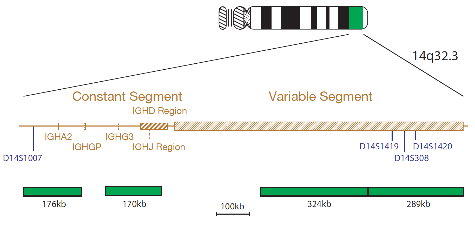
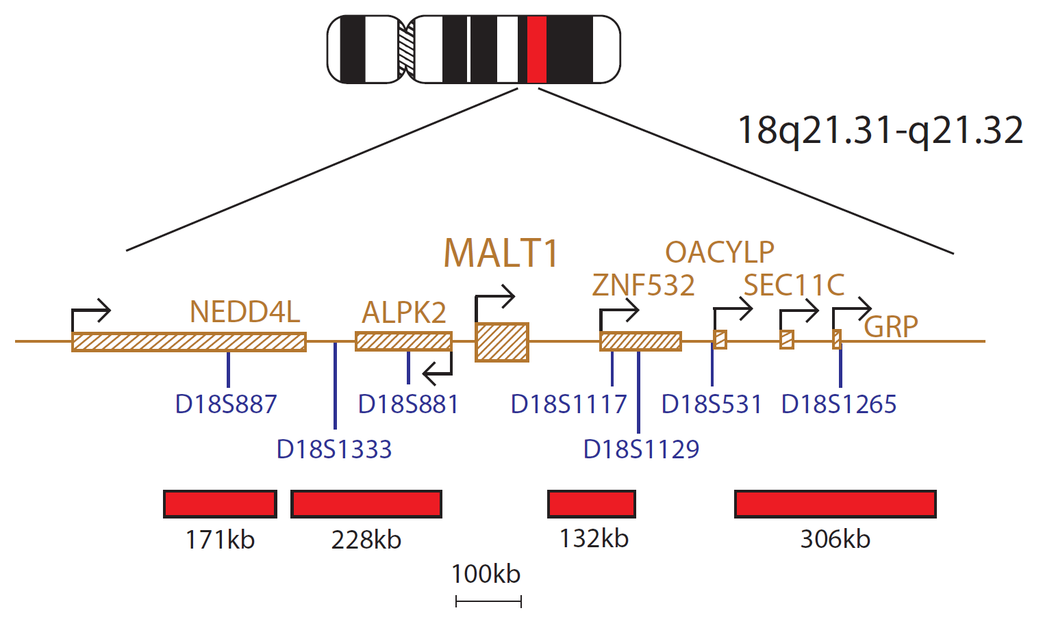
The IGH/MALT1 product consists of probes, labelled in green, covering the Constant, J, D and Variable segments of the IGH gene, and MALT1 probes, labelled in red. The MALT1 probe mix contains four probes, two 171kb, 228kb probes positioned centromeric to the MALT1 gene and two 132kb, 306kb probes positioned telomeric to the MALT1 gene.
The t(14;18)(q32;q21) involving the Immunoglobulin heavy chain locus (IGH) at 14q32 and the Mucosa Associated Lymphoid Tissue Lymphoma Translocation gene 1 (MALT1) at 18q21 is a recurrent abnormality in MALT lymphoma1.
Extranodal marginal zone B-cell lymphoma of the mucosa-associated lymphoid tissue is listed as a distinct clinicopathologic entity. MALT lymphomas comprise 7.6% of all non-Hodgkin lymphoma's (NHLs) and represents 1 of the 6 most common NHLs2. MALT lymphomas are characterised by a proliferation of neoplastic marginal zone-related cells that invade the epithelial structures to generate lymphoepithelial lesions and colonise reactive lymphoid follicles3. t(14;18)/IGH-MALT1- positive MALT lymphomas originate in sites such as the liver, skin, ocular adnexa, or salivary glands1. The molecular characteristics of the t(14;18) resemble those found in the t(14;18)/IGH-BCL2 (follicular lymphoma) and t(11;14)/MALT1-IGH (mantle cell lymphoma), suggesting that these translocations could be generated by common pathomechanisms involving illegitimate V(D)J-mediated recombination of IGH as well as non-homologous end joining (NHEJ) or alternative NHEJ repair pathways1.
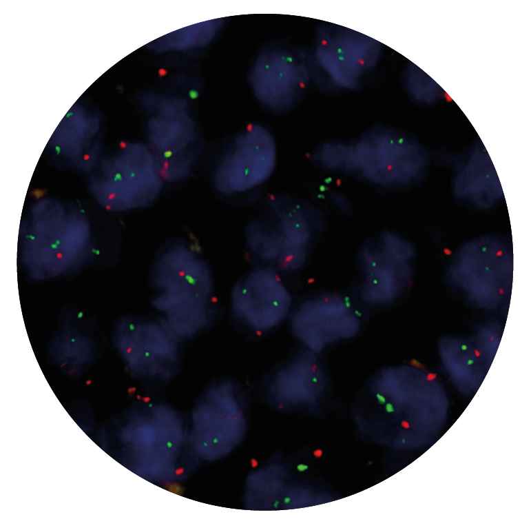
Microscope image
In vitro diagnostic (IVD)
→ English/Français/Italiano/Deutsch/Español
→ Polski
Research use only (RUO)
→ English
Find certificate of analysis documentation for our CytoCell FISH probes

Our lab has been using a wide range of CytoCell FISH probes for a number of years, and have been increasing this range all the time. The probes have clear bright signals and show good reproducibility. CytoCell provides fast delivery of catalogue probes, and are very responsive when we have any queries or problems with their products.

Bridget Manasse
Addenbrookes Hospital, Cambridge University Hosiptals NHS Foundation Trust, UK
In our hands, CytoCell FISH probes have proven to be of the highest quality with bright, easy to interpret signals, thus providing confidence in our results. OGT's customer support is outstanding, as their staff are extremely knowledgeable and truly care about their customers and their customers’ needs.

Jennie Thurston
Director of Cytogenetics, Carolinas Pathology Group, USA
I first came across CytoCell FISH probes in a previous lab I worked in and I was struck by the quality of the products. Since this time, I have been recommending and introducing CytoCell probes across all application areas — now they are the primary FISH probes used in our lab. They have an excellent range of products and their ready-to-use reagent format saves considerable time.

Elizabeth Benner
Medical Technologist, University of Arizona Health Network, USA
We have been working with CytoCell fish probes for two decades because of their excellent clarity and intensity regardless of the size of the probe. It is so clear and simple to detect.
Dr. Marina Djurisic
Head of Laboratory of Medical Genetics, Mother and Child Health Care Institute of Serbia “Dr Vukan Cupic”, Serbia
The quality and consistency of CytoCell’s probes means I can trust the results, and my clients get their results in a timely manner.

Dr. Theresa C. Brown
Director, Cytogenetics Laboratory, Hayward Genetics Center, Tulane University School of Medicine, USA
It was very important for us to have more consistent results with our probes — easy-to-read bright signals and a range of vial sizes, which is much more cost-effective.

Janet Cowan, PhD
Director of the Cytogenetics Laboratory, Tufts Medical Center, USA
Not only do CytoCell offer an extensive range of high-quality FISH probes, the customer support is also excellent — providing fast access to all the probes I need. The probes are highly consistent with bright signals allowing easy scoring of results.
Dr. Eric Crawford
Senior Director, Genetics Associates Inc., USA
The quality and reproducibility of results using the CytoCell kit has been vital in accurately detecting co-deletions in our glioma investigations. We now have a cost-effective test that we can rely on that is also easy to use and interpret. We've been consistently impressed with this kit - not to mention the support offered by OGT's customer service, and have completely transitioned over to CytoCell probes.
Gavin Cuthbert, FRCPath
Head of Cancer Cytogenetics, Northern Genetics Servce, Newcastle, UK
 Visit USA site
 Visit Canada site


