


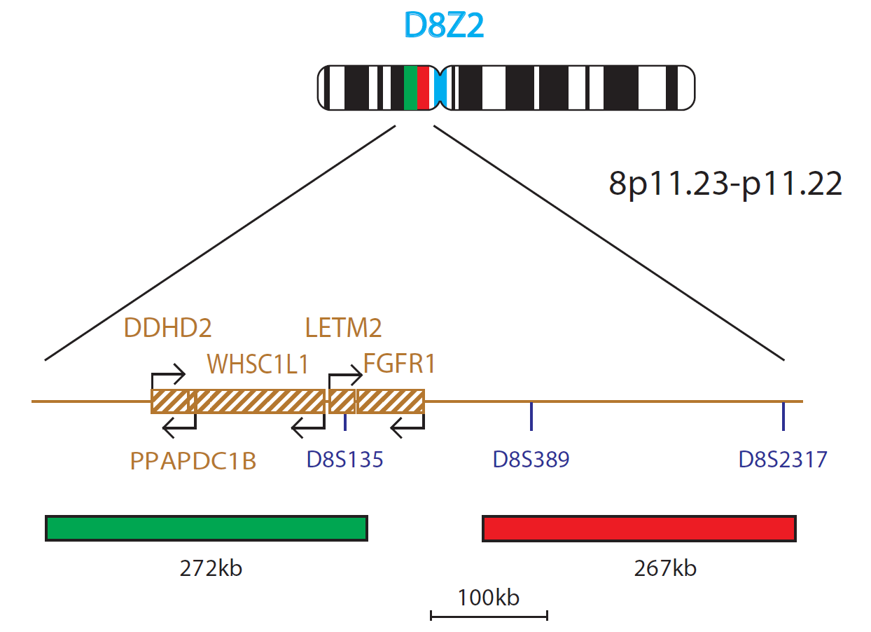
The FGFR1 Breakapart/Amplification probe consists of a green 272kb probe and a red 267kb probe, which are positioned on each side of the FGFR1 gene. The 8-centromere probe in blue acts as a control for chromosome 8.
Fibroblast Growth Factor Receptor 1 (FGFR1), located on chromosome 8, was the first gene to be shown to be amplified in some breast cancers1 and is also translocated in some patients with 8p11 myeloproliferative syndrome2.
Amplification of the gene is thought to be the causative factor in around 10% of breast tumours3 and has been shown to provide a poor prognosis to patients as over-expression of the gene product can lead to early relapse4.
In common with other genetic defects in lymphoproliferative disorders, like the BCR/ABL translocation in CML, the FGFR1 gene contains sequences that could code for a putative tyrosine kinase protein5,6, which has been implicated in myeloproliferative syndrome (also known as EMS or Stem cell leukaemia/lymphoma syndrome, SCLL), marking out the importance of this gene in both solid tumours and cancers of the blood. There are at least eight partner genes involved with FGFR1 translocations, including ZNF198 (the most common7), FOP, CEP110, BCR, HERV-K, FGFR10P2, TIF1 and MYO18A8.
CytoCell has developed a three-colour combined breakapart and amplification FISH probe for FGFR1 that can be used in either bone marrow samples from myeloproliferative syndrome (EMS) patients or tissue sections from patients where gene amplification may be suspected. In EMS, the breakapart strategy will show the split of one of the two fusion signals along with a blue centromere to enumerate chromosome 8. In patients that are being tested for amplifications of FGFR1, the non-translocated FGFR1 FISH probe will appear as a fusion signal and this will appear amplified. The chromosome 8 centromere probe will also act as a control probe in these cases.
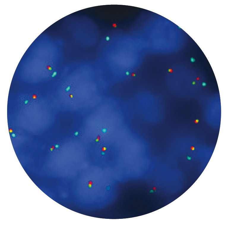
Microscope image
In vitro diagnostic (IVD)
→ English/Français/Italiano/Deutsch/Español
→ Polski
Research use only (RUO)
→ English
Find certificate of analysis documentation for our CytoCell FISH probes

Our lab has been using a wide range of CytoCell FISH probes for a number of years, and have been increasing this range all the time. The probes have clear bright signals and show good reproducibility. CytoCell provides fast delivery of catalogue probes, and are very responsive when we have any queries or problems with their products.

Bridget Manasse
Addenbrookes Hospital, Cambridge University Hosiptals NHS Foundation Trust, UK
In our hands, CytoCell FISH probes have proven to be of the highest quality with bright, easy to interpret signals, thus providing confidence in our results. OGT's customer support is outstanding, as their staff are extremely knowledgeable and truly care about their customers and their customers’ needs.

Jennie Thurston
Director of Cytogenetics, Carolinas Pathology Group, USA
I first came across CytoCell FISH probes in a previous lab I worked in and I was struck by the quality of the products. Since this time, I have been recommending and introducing CytoCell probes across all application areas — now they are the primary FISH probes used in our lab. They have an excellent range of products and their ready-to-use reagent format saves considerable time.

Elizabeth Benner
Medical Technologist, University of Arizona Health Network, USA
We have been working with CytoCell fish probes for two decades because of their excellent clarity and intensity regardless of the size of the probe. It is so clear and simple to detect.
Dr. Marina Djurisic
Head of Laboratory of Medical Genetics, Mother and Child Health Care Institute of Serbia “Dr Vukan Cupic”, Serbia
The quality and consistency of CytoCell’s probes means I can trust the results, and my clients get their results in a timely manner.

Dr. Theresa C. Brown
Director, Cytogenetics Laboratory, Hayward Genetics Center, Tulane University School of Medicine, USA
It was very important for us to have more consistent results with our probes — easy-to-read bright signals and a range of vial sizes, which is much more cost-effective.

Janet Cowan, PhD
Director of the Cytogenetics Laboratory, Tufts Medical Center, USA
Not only do CytoCell offer an extensive range of high-quality FISH probes, the customer support is also excellent — providing fast access to all the probes I need. The probes are highly consistent with bright signals allowing easy scoring of results.
Dr. Eric Crawford
Senior Director, Genetics Associates Inc., USA
The quality and reproducibility of results using the CytoCell kit has been vital in accurately detecting co-deletions in our glioma investigations. We now have a cost-effective test that we can rely on that is also easy to use and interpret. We've been consistently impressed with this kit - not to mention the support offered by OGT's customer service, and have completely transitioned over to CytoCell probes.
Gavin Cuthbert, FRCPath
Head of Cancer Cytogenetics, Northern Genetics Servce, Newcastle, UK
 Visit USA site
 Visit Canada site


