


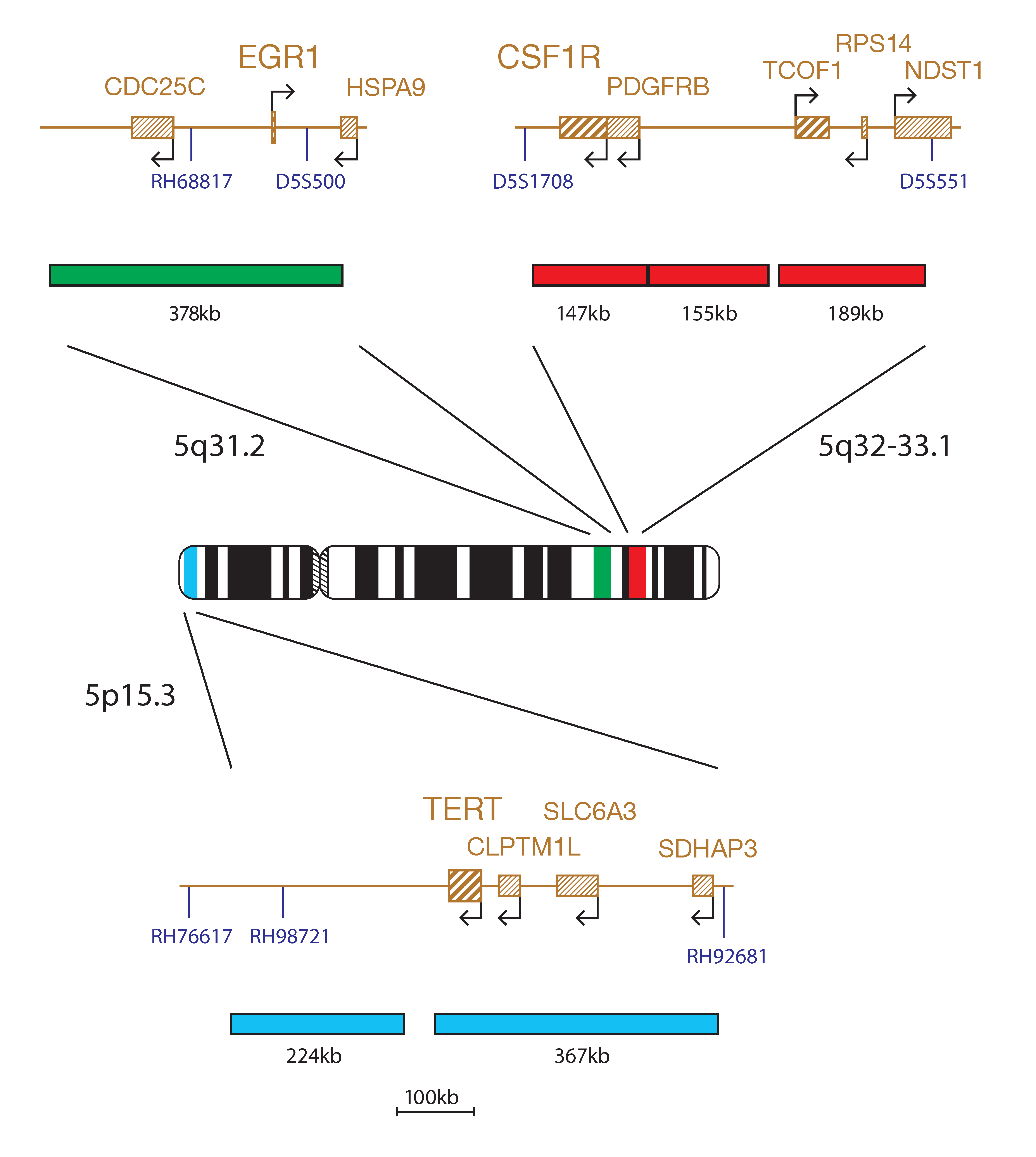
The Del(5q) Plus Tri-Colour Deletion Probe mix consists of three distinct probes. The green probe (378kb) covers the CDC25C and EGR1 genes, along with their flanking regions that include the RH68817 and D5S500 markers. The red probe set (147kb, 155kb and 189kb) locates between the D5S1708 and D5S551 markers and includes the CSF1R, PDGFRB, TCOF1 and RPS14 genes. The aqua probe set (224kb and 367kb) locates between the markers RH76617 and RH92681 and includes the genes TERT, CLPTM1L, SLC6A3 and SDHAP3.
Deletions of the long arm of chromosome 5 are one of the most common karyotypic abnormalities in myelodysplastic syndrome (MDS) and acute myeloid leukaemia (AML) with myelodysplasia related changes1,2.
A subset of MDS patients with del(5q) as a sole cytogenetic abnormality, or with a single additional abnormality not involving chromosome 7, have a consistent set of clinical features, termed the 5q- syndrome1. It is the only MDS subtype defined cytogenetically in the World Health Organization classification system. This clinical entity with <5% blasts has a more favourable prognosis. However, patients with del(5q) associated with other cytogenetic abnormalities, or with excess blasts, have an inferior survival2,3.
In contrast to de novo MDS, prognosis of AML with del(5q) is generally unfavourable, especially when seen as a part of complex karyotype4. Deletion of 5q is also commonly seen in treatment related t-MDS and t-AML where prognosis is particularly poor1.
Two chromosomal regions have been mapped on chromosome 5q in MDS and AML. One common deleted region, at 5q33, is associated with the 5q- syndrome. Another, more proximal region located at 5q31, has been linked to a more aggressive form of MDS and AML and is often accompanied by additional cytogenetics abnormalities and a poorer prognosis1,3,5.
The CytoCell Del(5q) Plus Tri-Colour Deletion Probe will detect deletions of EGR1 (early growth response 1), a tumour suppressor gene at 5q31. EGR1 has been shown to act through haploinsufficiency to initiate the development of MDS/AML6. The probe will also detect deletions of RPS14 (ribosomal protein S14) at 5q33.1, patients with MDS with del(5q) are haploinsufficient for RPS14 which leads to impairment of ribosome biogenesis and affects translation of genes and activation of proteins involved in differentiation and apoptosis4. The TERT (telomerase reverse transcriptase) gene probe at 5p15.3 will help distinguish cases with del(5q) from those with monosomy 5.
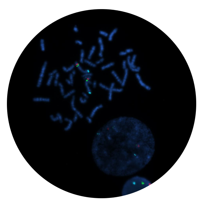
Microscope image
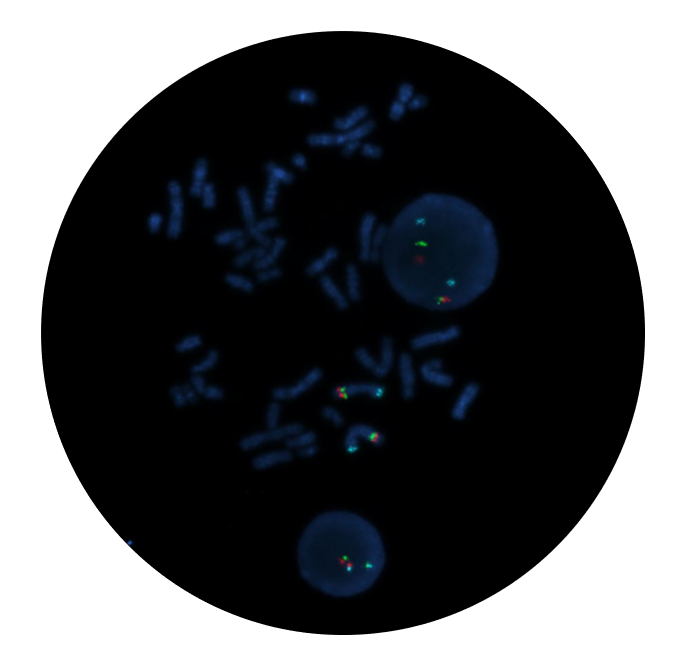
Microscope image
Find certificate of analysis documentation for our CytoCell FISH probes

Our lab has been using a wide range of CytoCell FISH probes for a number of years, and have been increasing this range all the time. The probes have clear bright signals and show good reproducibility. CytoCell provides fast delivery of catalogue probes, and are very responsive when we have any queries or problems with their products.

Bridget Manasse
Addenbrookes Hospital, Cambridge University Hosiptals NHS Foundation Trust, UK
In our hands, CytoCell FISH probes have proven to be of the highest quality with bright, easy to interpret signals, thus providing confidence in our results. OGT's customer support is outstanding, as their staff are extremely knowledgeable and truly care about their customers and their customers’ needs.

Jennie Thurston
Director of Cytogenetics, Carolinas Pathology Group, USA
I first came across CytoCell FISH probes in a previous lab I worked in and I was struck by the quality of the products. Since this time, I have been recommending and introducing CytoCell probes across all application areas — now they are the primary FISH probes used in our lab. They have an excellent range of products and their ready-to-use reagent format saves considerable time.

Elizabeth Benner
Medical Technologist, University of Arizona Health Network, USA
We have been working with CytoCell fish probes for two decades because of their excellent clarity and intensity regardless of the size of the probe. It is so clear and simple to detect.
Dr. Marina Djurisic
Head of Laboratory of Medical Genetics, Mother and Child Health Care Institute of Serbia “Dr Vukan Cupic”, Serbia
The quality and consistency of CytoCell’s probes means I can trust the results, and my clients get their results in a timely manner.

Dr. Theresa C. Brown
Director, Cytogenetics Laboratory, Hayward Genetics Center, Tulane University School of Medicine, USA
It was very important for us to have more consistent results with our probes — easy-to-read bright signals and a range of vial sizes, which is much more cost-effective.

Janet Cowan, PhD
Director of the Cytogenetics Laboratory, Tufts Medical Center, USA
Not only do CytoCell offer an extensive range of high-quality FISH probes, the customer support is also excellent — providing fast access to all the probes I need. The probes are highly consistent with bright signals allowing easy scoring of results.
Dr. Eric Crawford
Senior Director, Genetics Associates Inc., USA
The quality and reproducibility of results using the CytoCell kit has been vital in accurately detecting co-deletions in our glioma investigations. We now have a cost-effective test that we can rely on that is also easy to use and interpret. We've been consistently impressed with this kit - not to mention the support offered by OGT's customer service, and have completely transitioned over to CytoCell probes.
Gavin Cuthbert, FRCPath
Head of Cancer Cytogenetics, Northern Genetics Servce, Newcastle, UK
 Visit USA site
 Visit Canada site


