


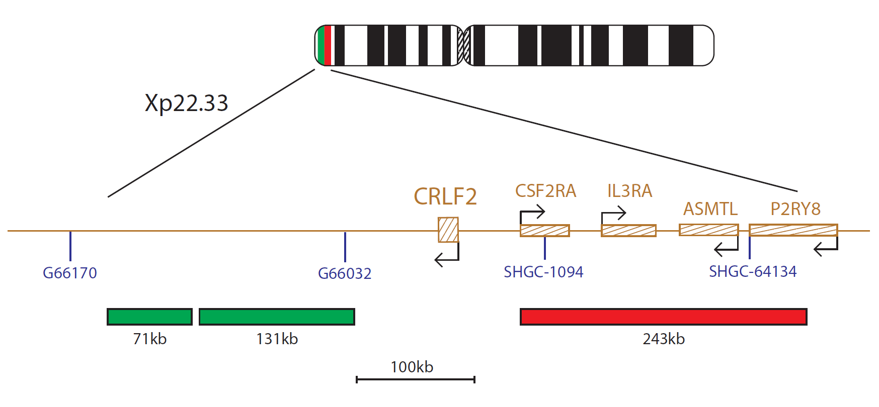
The CRLF2 Breakapart probe consists of a red 243kb probe, which is centromeric to the CRLF2 gene, and two green probes (71kb, 131kb), which are telomeric to CRLF2.
Overexpression of Cytokine Receptor-Like Factor 2 (CRLF2) has been shown to occur in Acute Lymphoblastic Leukaemia (ALL), particularly that associated with Down Syndrome (DS-ALL)1,2.
Overexpression of this protein has been associated with activation of the JAK/STAT5 pathway in transduced primary B-cell progenitors1,3 and various groups have attempted to characterise the biochemical consequences of these genetic lesions, with the goal of identifying targets for new therapies2,3.
CRLF2 overexpression has been shown to be caused by chromosomal rearrangements including the t(X;14) or t(Y;14) translocations or interstitial deletions of the pseudoautosomal region 1 (PAR1) of chromosomes X and Y. These place CRLF2 under control of an IGH enhancer1 or juxtapose the first non-coding exon of P2RY8 to the coding region of CRLF22, respectively. All of these CRLF2 rearrangements are cytogenetically cryptic and cannot be detected by conventional G-banded analysis1, making FISH a powerful detection tool for these abnormalities and an aid to furthering this research. Our CRLF2 Breakapart probe will detect rearrangements of the CRLF2 gene, whilst the P2RY8 Deletion probe allows detection of deletions between CRLF2 and P2RY8, causing the fusion gene.
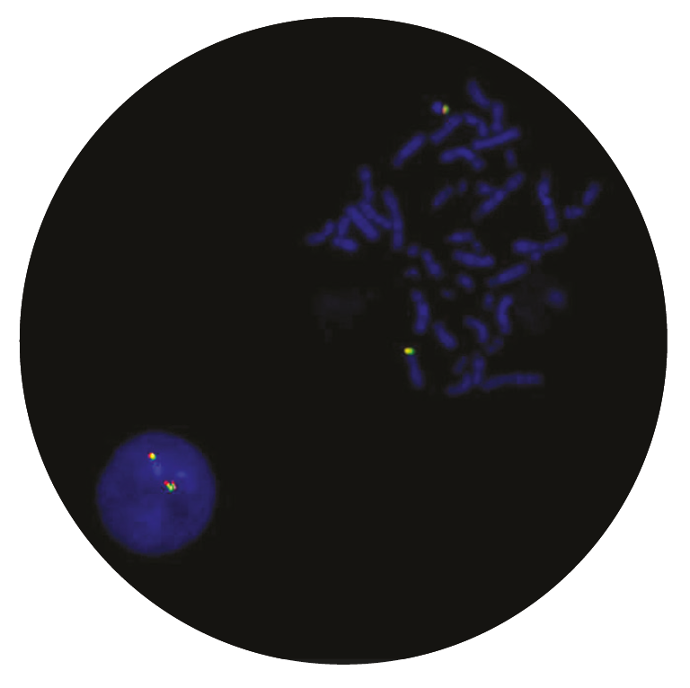
Microscope image
Research use only (RUO)
Find certificate of analysis documentation for our CytoCell FISH probes

Our lab has been using a wide range of CytoCell FISH probes for a number of years, and have been increasing this range all the time. The probes have clear bright signals and show good reproducibility. CytoCell provides fast delivery of catalogue probes, and are very responsive when we have any queries or problems with their products.

Bridget Manasse
Addenbrookes Hospital, Cambridge University Hosiptals NHS Foundation Trust, UK
In our hands, CytoCell FISH probes have proven to be of the highest quality with bright, easy to interpret signals, thus providing confidence in our results. OGT's customer support is outstanding, as their staff are extremely knowledgeable and truly care about their customers and their customers’ needs.

Jennie Thurston
Director of Cytogenetics, Carolinas Pathology Group, USA
I first came across CytoCell FISH probes in a previous lab I worked in and I was struck by the quality of the products. Since this time, I have been recommending and introducing CytoCell probes across all application areas — now they are the primary FISH probes used in our lab. They have an excellent range of products and their ready-to-use reagent format saves considerable time.

Elizabeth Benner
Medical Technologist, University of Arizona Health Network, USA
We have been working with CytoCell fish probes for two decades because of their excellent clarity and intensity regardless of the size of the probe. It is so clear and simple to detect.
Dr. Marina Djurisic
Head of Laboratory of Medical Genetics, Mother and Child Health Care Institute of Serbia “Dr Vukan Cupic”, Serbia
The quality and consistency of CytoCell’s probes means I can trust the results, and my clients get their results in a timely manner.

Dr. Theresa C. Brown
Director, Cytogenetics Laboratory, Hayward Genetics Center, Tulane University School of Medicine, USA
It was very important for us to have more consistent results with our probes — easy-to-read bright signals and a range of vial sizes, which is much more cost-effective.

Janet Cowan, PhD
Director of the Cytogenetics Laboratory, Tufts Medical Center, USA
Not only do CytoCell offer an extensive range of high-quality FISH probes, the customer support is also excellent — providing fast access to all the probes I need. The probes are highly consistent with bright signals allowing easy scoring of results.
Dr. Eric Crawford
Senior Director, Genetics Associates Inc., USA
The quality and reproducibility of results using the CytoCell kit has been vital in accurately detecting co-deletions in our glioma investigations. We now have a cost-effective test that we can rely on that is also easy to use and interpret. We've been consistently impressed with this kit - not to mention the support offered by OGT's customer service, and have completely transitioned over to CytoCell probes.
Gavin Cuthbert, FRCPath
Head of Cancer Cytogenetics, Northern Genetics Servce, Newcastle, UK
 Visit USA site
 Visit Canada site


