


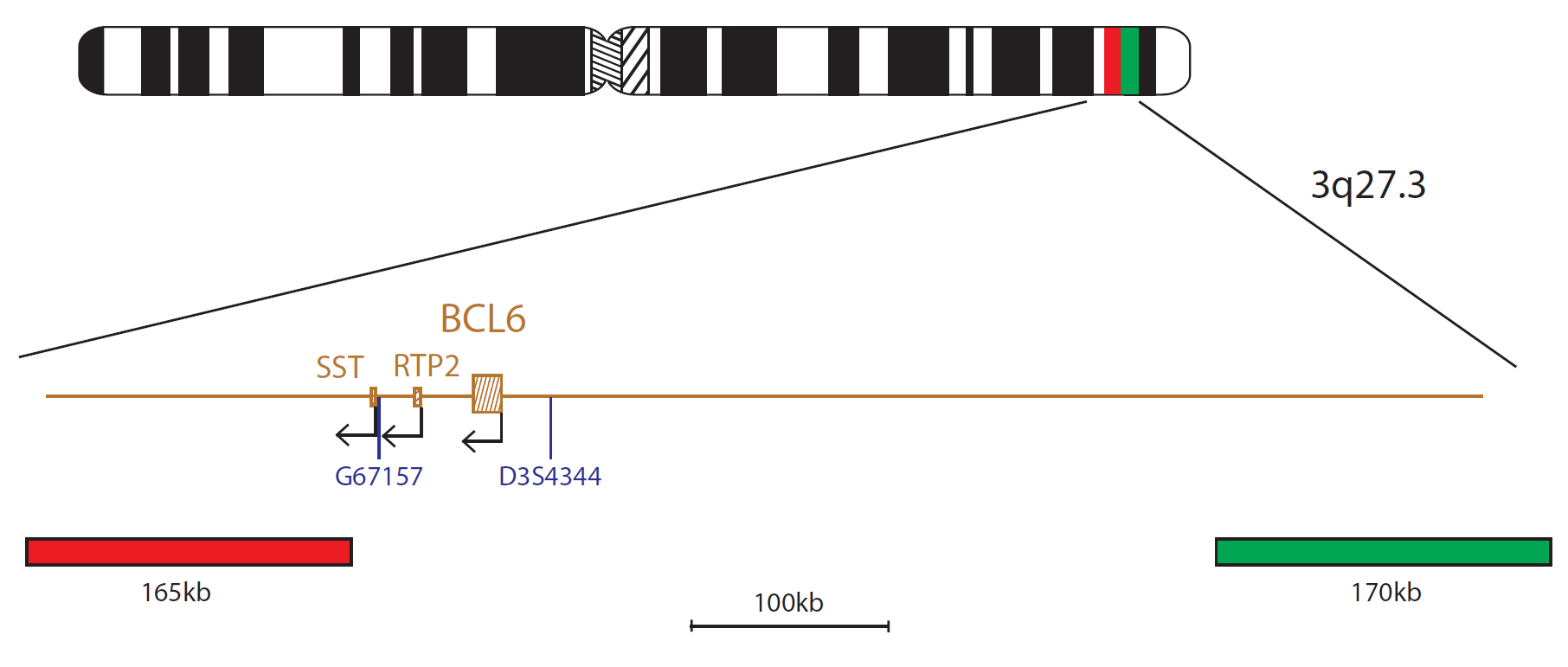
The BCL6 product consists of a 165kb probe, labelled in red, centromeric to the BCL6 gene and a green probe, covering a 170kb region telomeric to the BCL6 gene.
Chromosomal rearrangements involving the BCL6 (B-cell CLL/lymphoma 6) gene at 3q27 are recognised recurrent abnormalities commonly seen in patients with B-cell malignancy1.
BCL6 rearrangements are the most common chromosomal abnormalities seen in diffuse large B-cell lymphoma (DLBCL), occurring in up to 35% of cases2. They are also seen frequently in follicular lymphoma, where they occur in up to 15% of cases3. BCL6 is expressed in normal germinal centre B-cells and follicle helper T-cells. BCL6 translocations alter expression by promoter substitution and cause deregulated expression of normal BCL6 protein1,4.
Approximately 50% of BCL6 translocations will involve one of the three immunoglobulin loci (IGH, IGL or IGK); the remainder of translocations involve one of more than twenty different non-immunoglobulin genes5. Additionally, gains and amplifications of the BCL2 gene have also been reported in cases of B-cell lymphoma6.
The presence of concurrent BCL6 rearrangements with MYC and/or BCL2 rearrangements in patients with ‘dual-hit’ lymphoma has been shown to be associated with aggressive disease7.
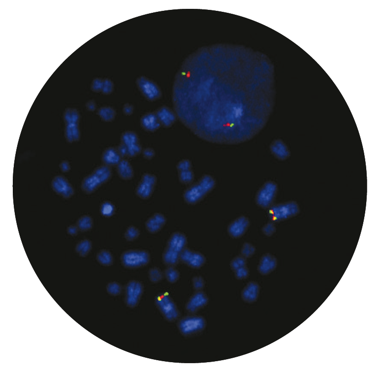
Microscope image
Find certificate of analysis documentation for our CytoCell FISH probes

Our lab has been using a wide range of CytoCell FISH probes for a number of years, and have been increasing this range all the time. The probes have clear bright signals and show good reproducibility. CytoCell provides fast delivery of catalogue probes, and are very responsive when we have any queries or problems with their products.

Bridget Manasse
Addenbrookes Hospital, Cambridge University Hosiptals NHS Foundation Trust, UK
In our hands, CytoCell FISH probes have proven to be of the highest quality with bright, easy to interpret signals, thus providing confidence in our results. OGT's customer support is outstanding, as their staff are extremely knowledgeable and truly care about their customers and their customers’ needs.

Jennie Thurston
Director of Cytogenetics, Carolinas Pathology Group, USA
I first came across CytoCell FISH probes in a previous lab I worked in and I was struck by the quality of the products. Since this time, I have been recommending and introducing CytoCell probes across all application areas — now they are the primary FISH probes used in our lab. They have an excellent range of products and their ready-to-use reagent format saves considerable time.

Elizabeth Benner
Medical Technologist, University of Arizona Health Network, USA
We have been working with CytoCell fish probes for two decades because of their excellent clarity and intensity regardless of the size of the probe. It is so clear and simple to detect.
Dr. Marina Djurisic
Head of Laboratory of Medical Genetics, Mother and Child Health Care Institute of Serbia “Dr Vukan Cupic”, Serbia
The quality and consistency of CytoCell’s probes means I can trust the results, and my clients get their results in a timely manner.

Dr. Theresa C. Brown
Director, Cytogenetics Laboratory, Hayward Genetics Center, Tulane University School of Medicine, USA
It was very important for us to have more consistent results with our probes — easy-to-read bright signals and a range of vial sizes, which is much more cost-effective.

Janet Cowan, PhD
Director of the Cytogenetics Laboratory, Tufts Medical Center, USA
Not only do CytoCell offer an extensive range of high-quality FISH probes, the customer support is also excellent — providing fast access to all the probes I need. The probes are highly consistent with bright signals allowing easy scoring of results.
Dr. Eric Crawford
Senior Director, Genetics Associates Inc., USA
The quality and reproducibility of results using the CytoCell kit has been vital in accurately detecting co-deletions in our glioma investigations. We now have a cost-effective test that we can rely on that is also easy to use and interpret. We've been consistently impressed with this kit - not to mention the support offered by OGT's customer service, and have completely transitioned over to CytoCell probes.
Gavin Cuthbert, FRCPath
Head of Cancer Cytogenetics, Northern Genetics Servce, Newcastle, UK
 Visit USA site
 Visit Canada site


