


Probe 1
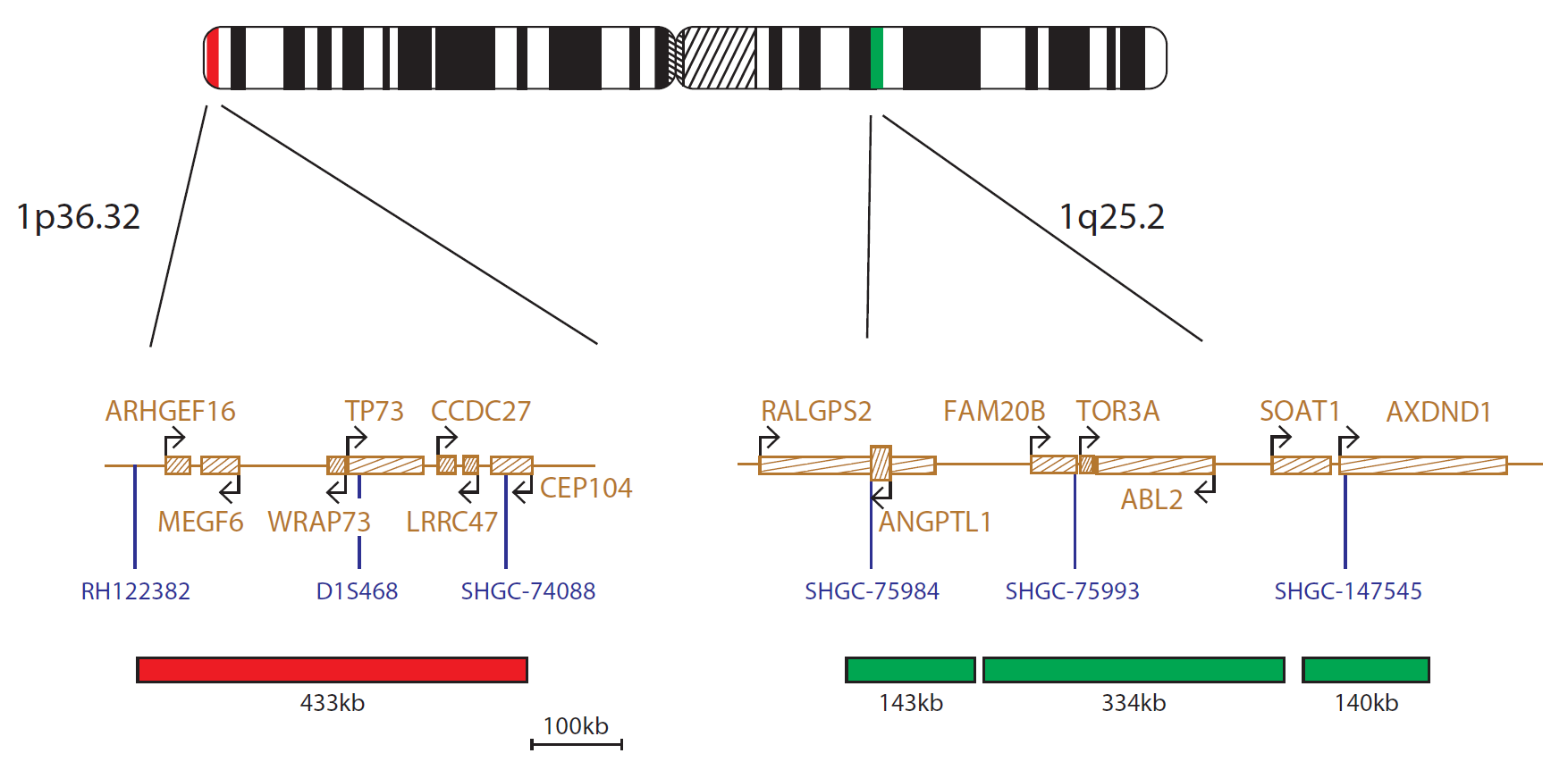
The 1p36.32 probe, labelled in red, is 433kb in size and covers the region between markers RH122382 and SHGC-74088. The 1q25.2 probe, labelled in green, consists of three probes (143kb, 334kb and 140kb) that cover regions including markers SHGC-75984 and SHGC-147545.
Probe 2
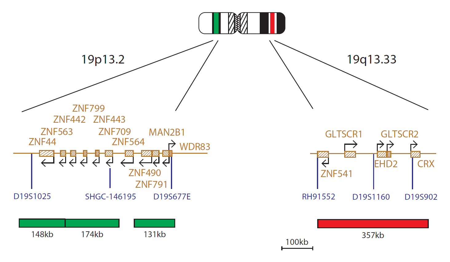
The 19p13.2 probe, labelled in green, consists of three probes (148kb, 174kb and 131kb) that cover regions including markers D19S1025 and D19S677E. The 19q13.33 probe, labelled in red, is 357kb in size and covers the region between markers RH91552 and D19S902.
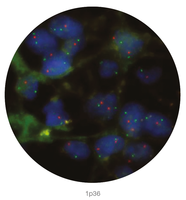
Microscope image of the 1p36 probe
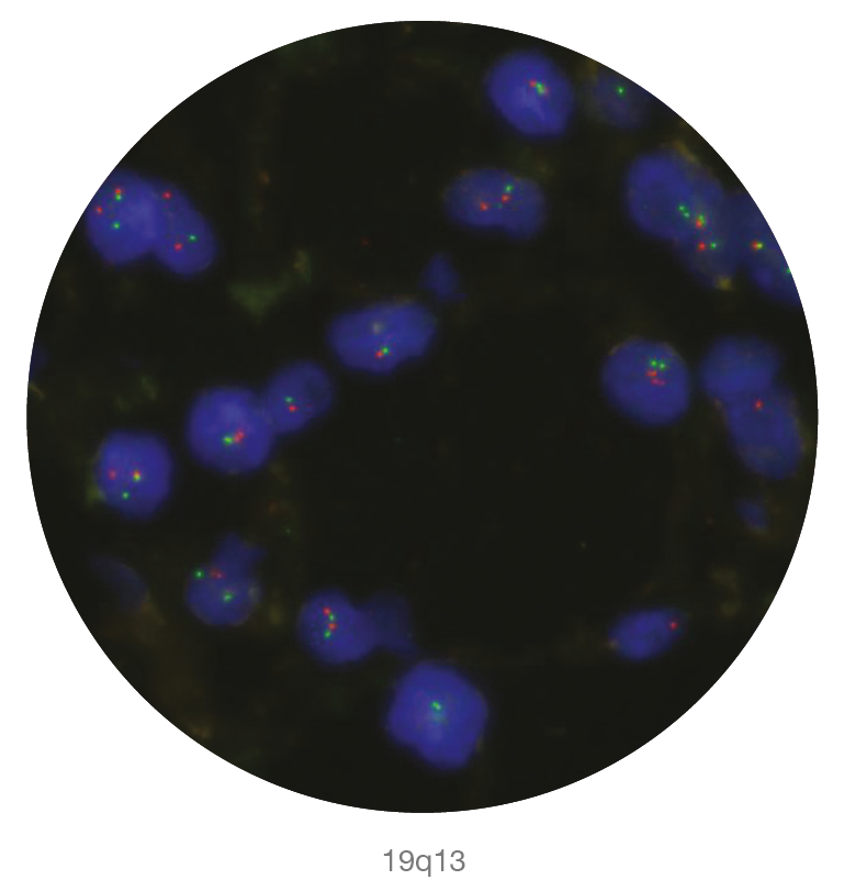
Microscope image of the 19q13 probe
Deletions of the 1p36.32 region including the TP73 (tumor protein 73) gene and deletions of the 19q13.33 region including the GLTSCR1 and GLTSCR2 (glioma tumor suppressor candidate region genes 1 and 2) genes are frequently reported in cases of glial tumours.
Astrocytomas and oligodendrogliomas are the most common gliomas that arise from glial cells. They make up about 40% of all CNS tumours1 and more than 60% of primary brain cancers2.
Concurrent losses, ‘co-deletion’, of the 1p36.32 and 19q13.33 regions are reported in approximately 80% of oligodendrogliomas, two-thirds of anaplastic oligodendrogliomas, as well as subsets of oligoastrocytomas and anaplastic oligoastrocytomas3,4; the majority of these losses have been shown to be mediated by the presence of an unbalanced t(1;19)(q10;p10) translocation. The presence of a 1p and 19q co-deletion is a strong prognostic factor in these diseases, where it is associated with improved prognosis and responsiveness to therapy5.
1p and 19q co-deletion has also been shown to occur in a subset of extraventricular neurocytomas, and may be associated with aggressive histology in these tumours6.
In vitro diagnostic (IVD)
→ English/Français/Italiano/Deutsch/Español
→ Polski
Research use only (RUO)
→ English
Find certificate of analysis documentation for our CytoCell FISH probes

Our lab has been using a wide range of CytoCell FISH probes for a number of years, and have been increasing this range all the time. The probes have clear bright signals and show good reproducibility. CytoCell provides fast delivery of catalogue probes, and are very responsive when we have any queries or problems with their products.

Bridget Manasse
Addenbrookes Hospital, Cambridge University Hosiptals NHS Foundation Trust, UK
In our hands, CytoCell FISH probes have proven to be of the highest quality with bright, easy to interpret signals, thus providing confidence in our results. OGT's customer support is outstanding, as their staff are extremely knowledgeable and truly care about their customers and their customers’ needs.

Jennie Thurston
Director of Cytogenetics, Carolinas Pathology Group, USA
I first came across CytoCell FISH probes in a previous lab I worked in and I was struck by the quality of the products. Since this time, I have been recommending and introducing CytoCell probes across all application areas — now they are the primary FISH probes used in our lab. They have an excellent range of products and their ready-to-use reagent format saves considerable time.

Elizabeth Benner
Medical Technologist, University of Arizona Health Network, USA
We have been working with CytoCell fish probes for two decades because of their excellent clarity and intensity regardless of the size of the probe. It is so clear and simple to detect.
Dr. Marina Djurisic
Head of Laboratory of Medical Genetics, Mother and Child Health Care Institute of Serbia “Dr Vukan Cupic”, Serbia
The quality and consistency of CytoCell’s probes means I can trust the results, and my clients get their results in a timely manner.

Dr. Theresa C. Brown
Director, Cytogenetics Laboratory, Hayward Genetics Center, Tulane University School of Medicine, USA
It was very important for us to have more consistent results with our probes — easy-to-read bright signals and a range of vial sizes, which is much more cost-effective.

Janet Cowan, PhD
Director of the Cytogenetics Laboratory, Tufts Medical Center, USA
Not only do CytoCell offer an extensive range of high-quality FISH probes, the customer support is also excellent — providing fast access to all the probes I need. The probes are highly consistent with bright signals allowing easy scoring of results.
Dr. Eric Crawford
Senior Director, Genetics Associates Inc., USA
The quality and reproducibility of results using the CytoCell kit has been vital in accurately detecting co-deletions in our glioma investigations. We now have a cost-effective test that we can rely on that is also easy to use and interpret. We've been consistently impressed with this kit - not to mention the support offered by OGT's customer service, and have completely transitioned over to CytoCell probes.
Gavin Cuthbert, FRCPath
Head of Cancer Cytogenetics, Northern Genetics Servce, Newcastle, UK
 Visit USA site
 Visit Canada site


