


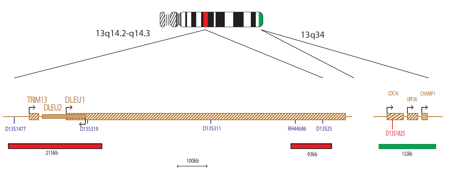
The 13q14.2-q14.3 probes, labelled in red, cover the D13S319 and D13S25 markers. The 13qter subtelomere specific probe (clone 163C9), labelled in green, allows identification of chromosome 13 and acts as a control probe.
Chromosome 13q abnormalities occur in 16-40% of multiple myeloma cases and are associated with poor prognosis1,2. A case study has shown that in 90% of patients, the 13q14 region was affected and 68% also showed involvement of the 13q21 region – the critical region in all but 8 patients was narrowed down to 13q143. Deletions affecting 13q14 are also the most frequent structural genetic abnormalities in B-cell Chronic Lymphocytic Leukaemia (B-CLL)4. This region is found to be heterozygously deleted in 30-60% and homozygously deleted in 10-20% of CLL patients5. Recently though, the survival rate has been shown to be similar for the two groups6. Two non-coding RNA genes, DLEU1 and DLEU2, and the genetic marker D13S319, span the pathogenic critical region of 13q147. DLEU1 is considered to be the most likely CLL-associated candidate tumour suppressor gene within the 13q14 region8. Subsequently, D13S319, located between the RB1 gene and D13S25 and within the DLEU1 locus, was found to be deleted in 44% of CLL cases9. It has also been postulated that a gene telomeric to the D13S319 region, encompassing D13S25, may be important in cases with hemizygous deletions and that this gene is a putative tumour suppressor gene10.
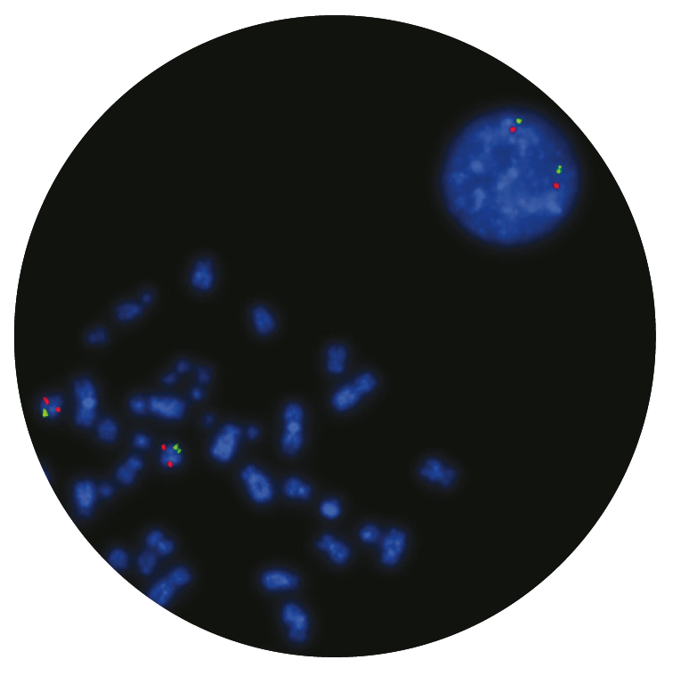
Microscope image
Find certificate of analysis documentation for our CytoCell FISH probes

Our lab has been using a wide range of CytoCell FISH probes for a number of years, and have been increasing this range all the time. The probes have clear bright signals and show good reproducibility. CytoCell provides fast delivery of catalogue probes, and are very responsive when we have any queries or problems with their products.

Bridget Manasse
Addenbrookes Hospital, Cambridge University Hosiptals NHS Foundation Trust, UK
In our hands, CytoCell FISH probes have proven to be of the highest quality with bright, easy to interpret signals, thus providing confidence in our results. OGT's customer support is outstanding, as their staff are extremely knowledgeable and truly care about their customers and their customers’ needs.

Jennie Thurston
Director of Cytogenetics, Carolinas Pathology Group, USA
I first came across CytoCell FISH probes in a previous lab I worked in and I was struck by the quality of the products. Since this time, I have been recommending and introducing CytoCell probes across all application areas — now they are the primary FISH probes used in our lab. They have an excellent range of products and their ready-to-use reagent format saves considerable time.

Elizabeth Benner
Medical Technologist, University of Arizona Health Network, USA
We have been working with CytoCell fish probes for two decades because of their excellent clarity and intensity regardless of the size of the probe. It is so clear and simple to detect.
Dr. Marina Djurisic
Head of Laboratory of Medical Genetics, Mother and Child Health Care Institute of Serbia “Dr Vukan Cupic”, Serbia
The quality and consistency of CytoCell’s probes means I can trust the results, and my clients get their results in a timely manner.

Dr. Theresa C. Brown
Director, Cytogenetics Laboratory, Hayward Genetics Center, Tulane University School of Medicine, USA
It was very important for us to have more consistent results with our probes — easy-to-read bright signals and a range of vial sizes, which is much more cost-effective.

Janet Cowan, PhD
Director of the Cytogenetics Laboratory, Tufts Medical Center, USA
Not only do CytoCell offer an extensive range of high-quality FISH probes, the customer support is also excellent — providing fast access to all the probes I need. The probes are highly consistent with bright signals allowing easy scoring of results.
Dr. Eric Crawford
Senior Director, Genetics Associates Inc., USA
The quality and reproducibility of results using the CytoCell kit has been vital in accurately detecting co-deletions in our glioma investigations. We now have a cost-effective test that we can rely on that is also easy to use and interpret. We've been consistently impressed with this kit - not to mention the support offered by OGT's customer service, and have completely transitioned over to CytoCell probes.
Gavin Cuthbert, FRCPath
Head of Cancer Cytogenetics, Northern Genetics Servce, Newcastle, UK
 Visit USA site
 Visit Canada site


