
In theory, any region of a chromosome can be a target for a FISH probe. CytoCell® probes may label chromosomes anywhere along the p or q arm: the subtelomere, the centromere, or any specific gene region in between.
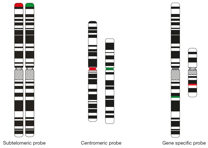
Figure 1 (above): Illustration of CytoCell subtelomeric, centromeric and gene specific FISH probes.
An example of the convention at OGT for assignment of a fluorophore* can be viewed with the FDA-cleared probe for AML/MDS: P53 (TP53) Deletion (USA-LPH 017):
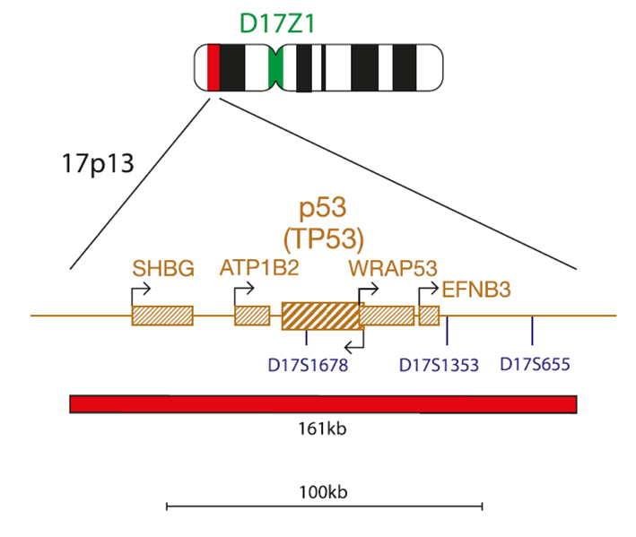
Figure 2 (above): CytoCell chromomap for P53 (TP53) Deletion
* There are some exceptions to this convention, please consult the IFU for probe map and full details.
Using the MLL (KMT2A) Breakapart probe (USA-LPH 013) probe as an example:
MLL (KMT2A) Breakapart
Breakapart = Probe design / signal pattern, one fluorophore for 3’ end and a different color fluorophore for 5’ end
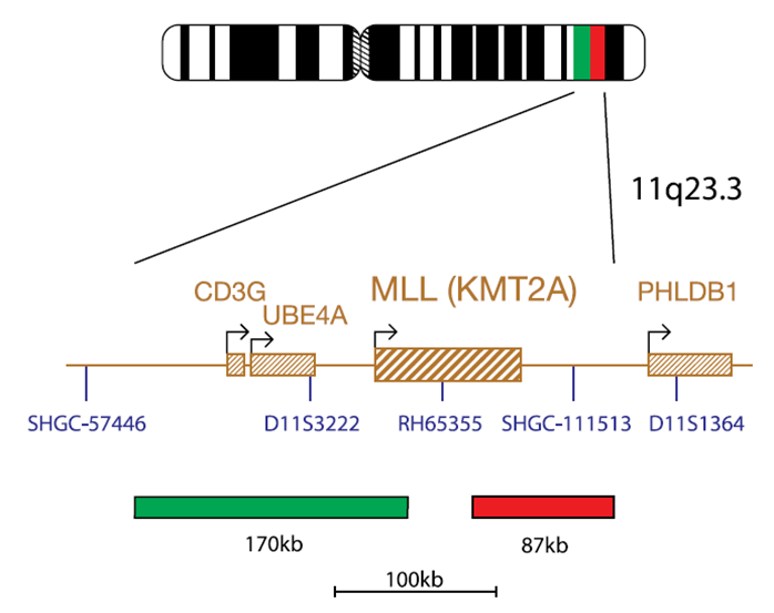 Figure 3 (above): CytoCell chromomap for MLL (KMT2A) Breakapart.
Probe maps on the OGT website have been updated to include information on gene orientation to allow the 3’ or 5’ positions of the probes to be seen in relation to the gene, or region, of interest. The arrows on the genes indicate the direction of transcription. Those with an arrow above, pointing to the right, are located on the plus, or sense, strand of the DNA. Those with an arrow below the gene, pointing to the left, are on the minus, antisense strand. In both cases, the 5’ end is that with the arrow as genes are transcribed in a 5’ to 3’ direction.
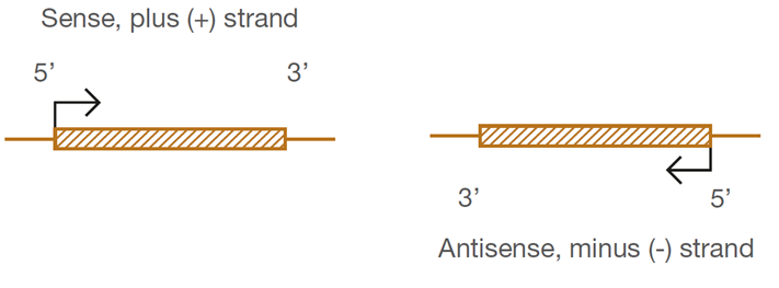
Figure 4 (above): Arrows on genes in chromomaps indicate the direction of transcription.

 Visit International site
 Visit Canada site