


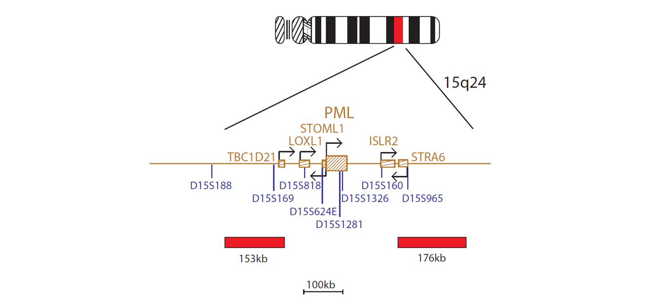
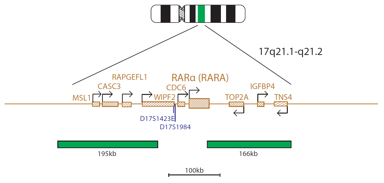
The PML probe mix, labelled in red, consists of a 153kb probe centromeric to the PML gene that covers the marker D15S169 and a 176kb probe telomeric to the PML gene that covers the marker D15S965. The RARα (RARA) probe mix, labelled in green, consists of a 195kb probe centromeric to the RARα (RARA) gene that spans the CASC3 gene and a 166kb probe that covers the telomeric end of the RARα (RARA) gene as well as the TOP2A, IGFBP4 and TNS4 genes.
The PML (promyelocytic leukaemia) gene is located at 15q24.1 and the RARA (retinoic acid receptor alpha) gene is located at 17q21.2. The translocation t(15;17)(q24;q21) gives rise to the PML::RARA fusion gene and is the diagnostic hallmark of acute promyelocytic leukaemia (APL).
This FAST PML/RARα FISH probe allows rapid detection of the rearrangement, with only one hour of hybridisation required.
The PML::RARA fusion gene is created by the t(15;17)(q24;q21) translocation, found in more than 90% of cases of APL, a leukaemia that comprises 5-8% of cases of acute myeloid leukaemia (AML)1,2. In a subset of cases, variant RARA translocations can be observed. Known fusion partners include NPM1 at 5q35, NUMA1 at 11q13, ZBTB16 (PLZF) at 11q23, STAT5B at 17q21, PRKAR1A at 17q24, FIP1L1 at 4q12 and BCOR at Xp113,4,5.
PML and RARA have both been implicated in normal haematopoiesis. PML possesses growth suppressor and proapoptotic activity whereas RARA is a transcription factor that mediates the effect of retinoic acid at specific response elements6. PML::RARA fusion protein behaves as an altered retinoic acid receptor with an ability of transmitting oncogenic signaling7.
Immediate treatment of APL patients is critical, due to fatal coagulation disorders and life-threatening haemorrhage in diagnosis. Prior to the introduction of all-trans-retinoic-acid (ATRA) and arsenic trioxide (ATO) in APL treatment protocols, the disease had a poor prognosis; however, since the introduction of these therapies, the overall survival rate has improved dramatically, with nearly 90%5 of patients cured. Patients with variant RARA translocations show variable sensitivity to treatment, with some patients showing resistance to treatment protocols3,5. It is therefore important to differentiate between APL patients with PML::RARA fusion and those patients with variant RARA translocations.

Microscope image
The CytoCell® FAST PML/RARα (RARA) Translocation, Dual Fusion Probe is a qualitative, non-automated, fluorescence in situ hybridisation (FISH) test used to detect chromosomal rearrangements between the 15q24 region on chromosome 15 and the 17q21.1-q21.2 region on chromosome 17 in Carnoy’s solution (3:1 methanol/acetic acid) fixed haematologically-derived cell suspensions from patients with confirmed or suspected acute myeloid leukaemia (AML).
This device is designed as an adjunct to other clinical and histopathological tests in recognised diagnostic and clinical care pathways, where knowledge of PML::RARA translocation status would be important for clinical management.
This device is designed to detect rearrangements with breakpoints in the region covered by the red and green clones in this probe set, which includes the PML and RARA regions. Breakpoints outside this region, or variant rearrangements wholly contained within this region, may not be detected with this device.
This device is not intended for: use as a stand-alone diagnostic, use as a companion diagnostic, prenatal testing, population-based screening, near-patient testing, or self-testing.
This device has not been validated for sample types, disease types, or purposes outside of those stated in the intended purpose.
It is intended as an adjunct to other diagnostic laboratory tests and therapeutic action should not be initiated on the basis of the FISH result alone.
Reporting and interpretation of FISH results should be performed by suitably qualified staff, consistent with professional standards of practice, and should take into consideration other relevant test results, clinical and diagnostic information.
This device is intended for laboratory professional use only.
Failure to adhere to the protocol may affect the performance and lead to false positive/negative results.
In our hands, CytoCell FISH probes, including the ROS1 Proximal and ROS1 Distal probes, have proven to be of the highest quality with bright, easy to interpret signals, thus providing confidence in our results. OGT's customer support is outstanding, as their staff are extremely knowledgeable and truly care about their customers and their customers’ needs.

Jennie Thurston
Director of Cytogenetics, Carolinas Pathology Group, USA

 Visit USA site
 Visit Canada site


