
Want to download this as a PDF? Download now
S Chatters, F Partheniou, A Hobbs, G Fonseka, A Castro-Justo, A Gocza-Blasko, K Mak-Hannah, R Frodsham, M Lawrie
Fluorescence in situ hybridisation (FISH) analysis is the ‘gold-standard’ method for the detection of balanced and unbalanced chromosomal rearrangements plus gains and deletions in neoplastic specimens. Standard FISH protocols incorporate an overnight hybridisation step; however, shorter hybridisations are sometimes desired as a result of laboratory operational requirements. The CytoCell® FISH probe range from OGT delivers bright, clear and precise signals when hybridised overnight. The aim of this project was to determine whether the hybridisation times for standard CytoCell ‘off-the-shelf’ CE-marked IVD probes could be reduced without detrimental effect on probe performance.
Ten CytoCell Haematology CE-marked IVD probe kits were selected for analysis, as shown in Table 1, below.
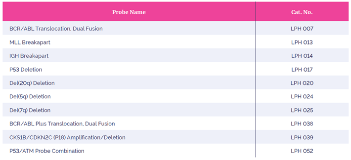 Table 1: Probe selection
Following the standard CytoCell protocol, (Table 2), four replicates of each probe set were hybridised to Carnoy’s-fixed peripheral blood lymphocyte and bone marrow samples and each replicate hybridised for five differing times: one hour, two hours, three hours, four hours and overnight (sixteen hours), making a total of 160 separate hybridisations.
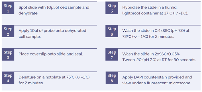 Table 2: Outline of Standard CytoCell FISH Procedure.
Each of the hybridisation replicates were analysed and assessed for individual probe component intensity and sensitivity by two independent analysts, scoring a target number of one hundred interphase nuclei each, according to CytoCell standard QC procedure, and validated against individual probe intensity measured using MetaSystems® Isis and Metafer image analysis software.
The independent scoring and intensity data were normalised against the data obtained for standard overnight hybridisation. This enabled relative sensitivity and intensity relative values to be calculated.
Absolute intensity, by pixel, and eye intensity values were combined to give a signal intensity index for each probe set. These indices were analysed by fluorophore colour and sample type, using a standard overnight hybridisation as baseline to give a measure of any reduction in signal intensity. Sensitivity data were normalised in the same manner, giving an indication of the probe sensitivity, alongside standard deviation values. These results are shown in Tables 2 and 3, and Figures 2 and 3.
At a hybridisation of four hours, the intensity and sensitivity indices for all sets of data are above 0.8 – the cut-off for CytoCell QC analysis. This indicates the presence of bright easily-scored signals with little dropout for all fluorophore types in all cell types analysed. Furthermore, many of the indices for hybridisation times below four hours are above or approaching the 0.8 value, which still indicates presence of strong bright signals, confirming the robustness of CytoCell probes when used with shorter hybridisation times. It is of note that even with a reduction of signal intensity below that of the CytoCell QC cut-off, signals remained bright and scoreable.
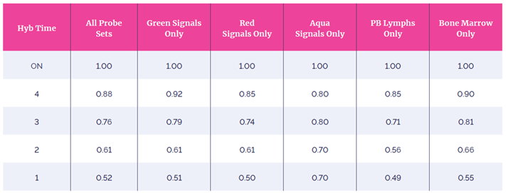 Table 3: Mean Intensity Index.
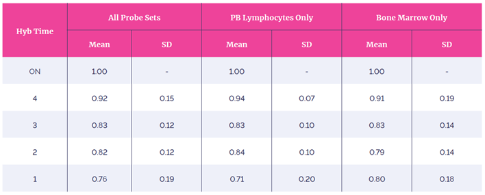 Table 4: Sensitivity Index.
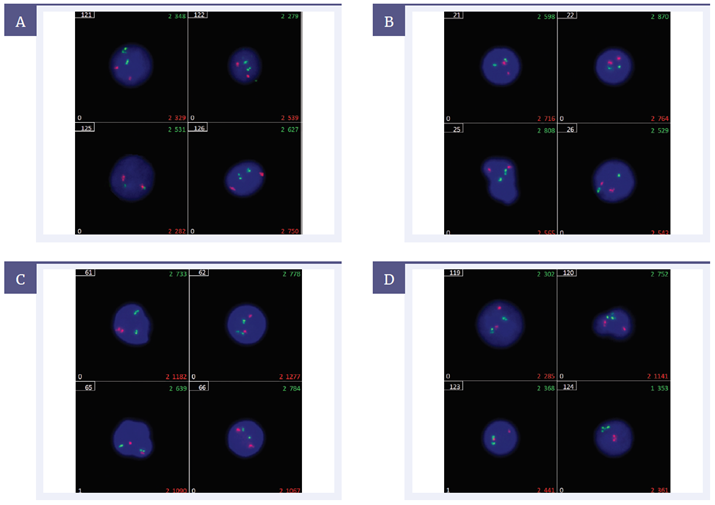 Figure 1: Comparison of hybridisation results. LPH007 probe set at [A] overnight, [B] four hours, [C] three hours, [D] one hour. Output from Metafer at 60x.
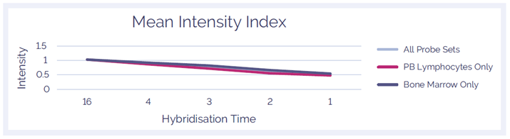 Figure 2: Mean Intensity Index
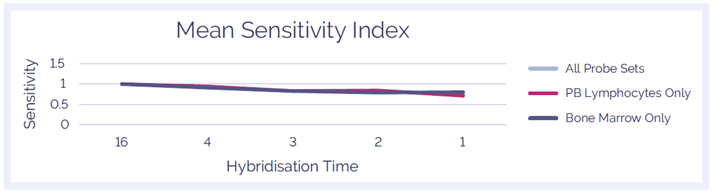 Figure 3: Mean Sensitivity Index
These data show that, for a range of probe and specimen types, the hybridisation time for standard CE IVD-marked CytoCell probes may be shortened from overnight to four hours or fewer, whilst retaining excellent signal sensitivity and intensity. This gives laboratories the flexibility to validate and use off-the-shelf CytoCell probes with reduced hybridisation times, facilitating same-day reporting, should this be required.
IVD: For in vitro diagnostic use. Some products may not be available in your region.
 Visit USA site
 Visit Canada site
