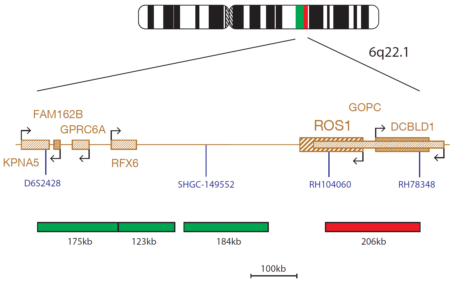
The ROS1 Plus probe consists of 3 probes (175kb, 123kb, 171kb), labelled in green, situated proximal to the ROS1 gene and including markers D6S2428 and SHGC-149552 and a red probe (206kb) covering the distal part of the ROS1 gene and a region up to RH78348.
The ROS1 (ROS proto-oncogene 1, receptor tyrosine kinase) gene at 6q22 is an ALK (anaplastic lymphoma receptor tyrosine kinase) gene paralogue which encodes a type I integral membrane protein with tyrosine kinase activity1.
ROS1 rearrangements define a molecular subset of non-small cell lung cancer (NSCLC) and are seen in approximately 2% of patients with NSCLC2. A number of partner genes have been identified, including SLC34A2, CD74 and SDC43. It has been shown that these ROS1 fusions activate the pSTAT3, PI3K/AKT/mTOR and SHP-2 phosphatase pathways4,5.
NSCLC patients with ROS1 rearrangements have been shown to respond to treatment with ALK/MET tyrosine kinase inhibitors, such as crizotinib6.
ROS1 rearrangements with the GOPC (golgi associated PDZ and coiled-coil motif containing) gene fusion partner were originally reported in glioblastoma, but have now also been detected on cholangiocarcinoma, ovarian cancer and NSCLC patient samples7-9.
The ROS1 Plus design covers the ROS1 region and the region deleted in ROS1-GOPC fusions.
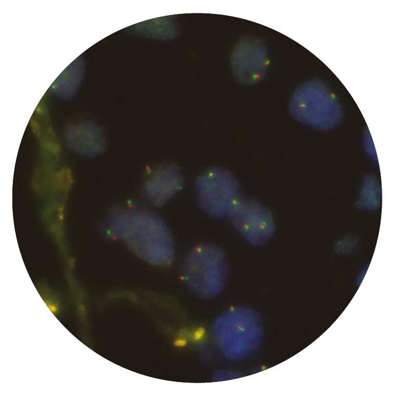
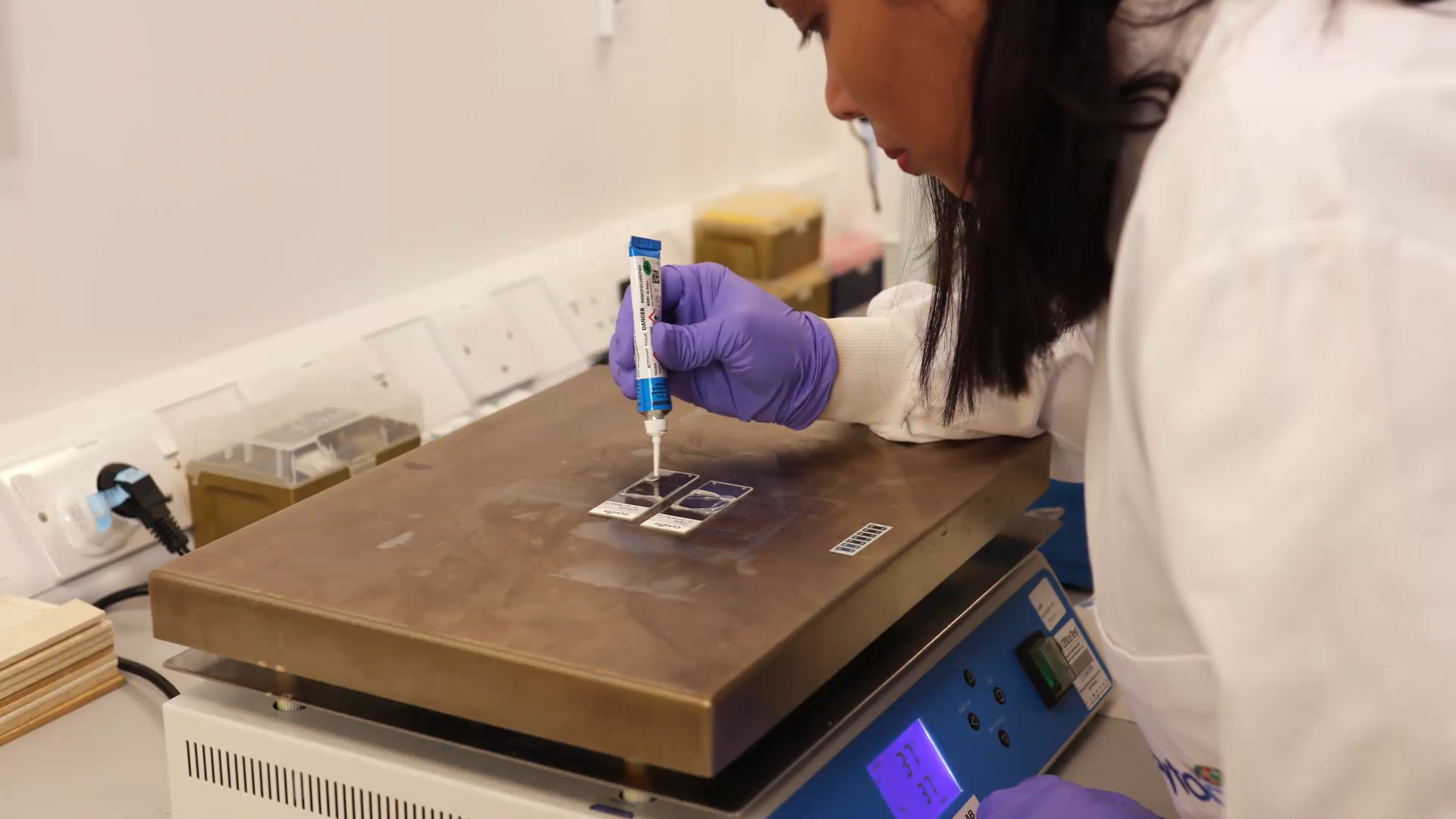
In our hands, CytoCell FISH probes, including the ROS1 Proximal and ROS1 Distal probes, have proven to be of the highest quality with bright, easy to interpret signals, thus providing confidence in our results. OGT's customer support is outstanding, as their staff are extremely knowledgeable and truly care about their customers and their customers’ needs.

Jennie Thurston
Director of Cytogenetics, Carolinas Pathology Group, USA

 Visit USA site
 Visit Canada site