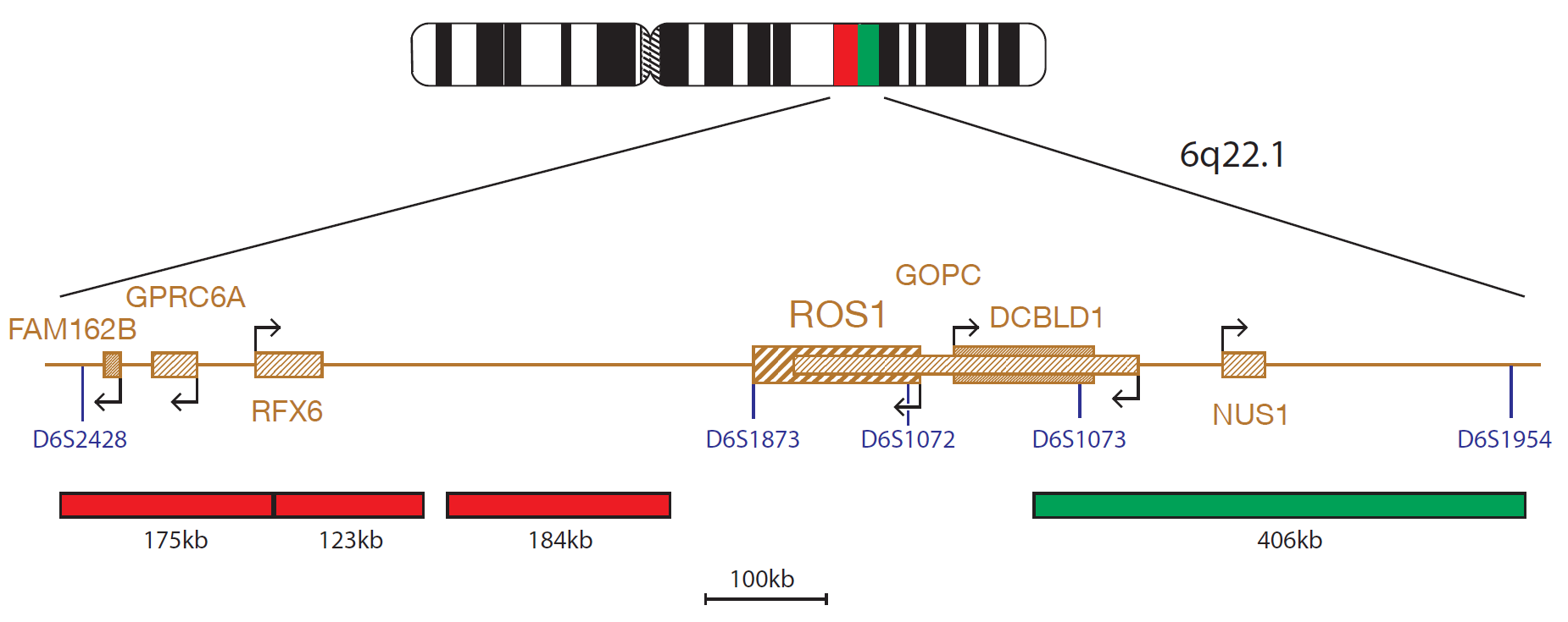
The ROS1 Breakapart probe consists of a green 406kb probe and two red 299kb and 171kb probes, which are positioned on each side of the ROS1 gene.
The receptor tyrosine kinase c-ros oncogene 1 (ROS1) is an orphan receptor tyrosine kinase of the insulin receptor family and is located at 6q221. ROS1 consists of a large extracellular domain that is composed of six fibronectin repeats, transmembrane domain, and an intracellular kinase domain. While the function of ROS1 is undefined, it has been shown to play an important role in the differentiation of epididymal epithelium2.
ROS1 rearrangements have been reported to define a molecular subset of Non Small-Cell Lung Cancer (NSCLC)3. Rearrangements of ROS1 with fusion partners SLC34A2 (solute carrier family 34 (sodium phosphate), member 2)4 and CD74 (Cluster of Differentiation 74), have been shown to occur in less than 2% of patients with NSCLC3. Patients harbouring ROS1 rearrangement have been shown to respond to treatment with ALK/MET tyrosine kinase inhibitors, such as Xalkori® (crizotinib)5,6.
ROS1 rearrangements with the fusion partner GOPC (golgi-associated PDZ and coiled-coil motif containing) have also been implicated in Glioblastoma and Cholangiocarcinoma5,7. ROS1 Fusions activate the pSTAT3, PI3K/AKT/mTOR and the SHP-2 phosphatase pathways8,9.
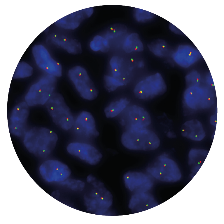
In our hands, CytoCell FISH probes, including the ROS1 Proximal and ROS1 Distal probes, have proven to be of the highest quality with bright, easy to interpret signals, thus providing confidence in our results. OGT's customer support is outstanding, as their staff are extremely knowledgeable and truly care about their customers and their customers’ needs.

Jennie Thurston
Director of Cytogenetics, Carolinas Pathology Group, USA

 Visit USA site
 Visit Canada site