


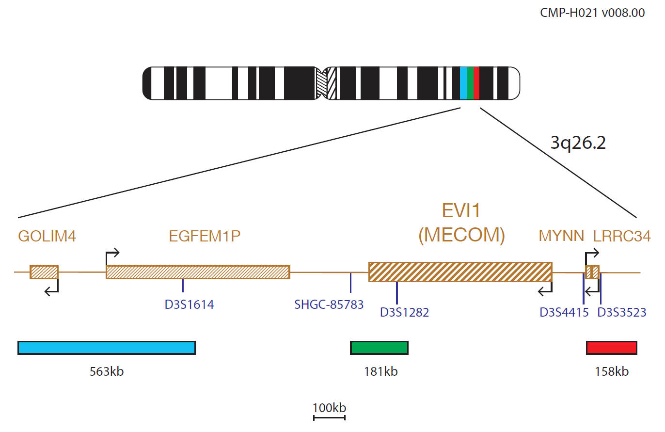
The red component of the EVI1 probe mix consists of a 158kb probe telomeric to the D3S4415 marker and includes the LRRC34 gene. The green component covers a 181kb region that includes the centromeric part of the EVI1 (MECOM) gene and beyond marker D3S1282. The aqua component covers a 563kb region centromeric to the EVI1 gene, that includes the D3S1614 marker.
The MECOM (MDS1 and EVI1 complex locus) oncogene at 3q26.2 is often rearranged in haematological malignancies of myeloid origin, including myelodysplastic neoplasms (MDS) and acute myeloid leukaemia with MECOM rearrangement (AML). Its expression in neoplastic myeloid cells disrupts myeloid differentiation, cell cycle regulation, and cell signalling pathways1.
This deregulated expression is often due to a chromosomal rearrangement involving 3q26.2, with the two most common (~40%) aberrations being the t(3;3)(q21;q26.2) and inv(3)(q21q26.2)1. More than 30 additional 3q26.2 rearrangements have been described, most of them characterised at the molecular level1.
The breakpoints for the translocations and inversions vary considerably. MECOM rearrangements are very heterogeneous and may be difficult to detect by conventional cytogenetics, making FISH a useful tool for their detection. Variant t(3;v)(q26.2;v) breakpoint regions can extend from 3′ proximal of MECOM to 5′ distal of the MDS1-EVI1 promoter, covered by the green probe. Therefore, the expected signal pattern for these translocations varies dependent on the breakpoint position2.Testing for MECOM rearrangements is recommended in both MDS and AML3.
AML with MECOM rearrangement is an aggressive disease with short survival irrespective of the blast percentage, with no difference in outcome between cases with inv(3)/t(3;3) as compared to MECOM rearrangements with other partners1. MDS risk-stratification incorporates variables such as age, severity of cytopenias, and cytogenetic findings1.
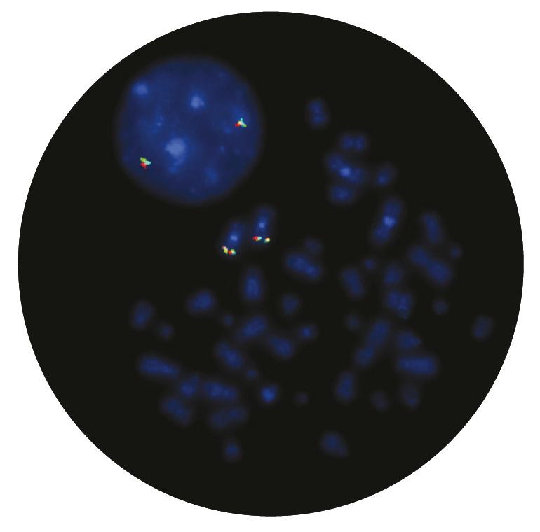
Microscope image
The CytoCell® EVI1 (MECOM) Breakapart Probe is a qualitative, non-automated, fluorescence in situ hybridisation (FISH) test used to detect chromosomal rearrangements involving the 3q26.2 region on chromosome 3 in Carnoy’s solution (3:1 methanol/acetic acid) fixed haematologically-derived cell suspensions from patients with confirmed or suspected acute myeloid leukaemia with MECOM rearrangement (AML) or myelodysplastic neoplasms (MDS).
This device is designed as an adjunct to other clinical and histopathological tests in recognised diagnostic and clinical care pathways, where knowledge of MECOM rearrangement status would be important for clinical management.
This device is designed to detect rearrangements with breakpoints in the region bounded by the red, green and aqua clones in this probe set, which includes the MECOM region (green probe), a region telomeric to the MECOM gene (red probe), and a region centromeric to the MECOM gene (aqua probe). Breakpoints outside these regions or variant rearrangements wholly contained within this region may not be detected with this device.
This device is not intended for: use as a stand-alone diagnostic, use as a companion diagnostic, prenatal testing, population-based screening, near-patient testing, or self-testing.
This device has not been validated for sample types, disease types, or purposes outside of those stated in the intended purpose.
It is intended as an adjunct to other diagnostic laboratory tests and therapeutic action should not be initiated on the basis of the FISH result alone.
Reporting and interpretation of FISH results should be performed by suitably qualified staff, consistent with professional standards of practice, and should take into consideration other relevant test results, clinical and diagnostic information.
This device is intended for laboratory professional use only.
Failure to adhere to the protocol may affect the performance and lead to false positive/negative results.
I first came across CytoCell FISH probes in a previous lab I worked in and I was struck by the quality of the products. Since this time, I have been recommending and introducing CytoCell probes across all application areas — now they are the primary FISH probes used in our lab. They have an excellent range of products and their ready-to-use reagent format saves considerable time.

Elizabeth Benner
Medical Technologist, University of Arizona Health Network, USA

 Visit USA site
 Visit Canada site


